

Description of appearance of ecchymosis on an arm, simultaneously with a classical leukocytoclastic vasculitis, the proposal of alternative utilities of measuring blood pressure, and the study of side effects to that measure.
PatientCase 80-year-old male came to ER with dyspnea, heart failure, predialysis renal failure with hyperkalemia and hemodynamic instability. During his stay he developed a skin lesion that looks like palpable purpura, from the lower limit of the blood pressure cuff to the distal area of the hand that not disappeared with vitropression, and pruritus. During admission the arm injury was extended to all members, both upper and lower.
ResultsThe study concluded with diagnosis of leukocytoclastic vasculitis given the presence of eosinophils, that which suggested probable drug etiology to an antibiotic that had been taken since seven days prior to admission to ER.
DiscussionThe need for serial monitoring of blood pressure, and the duration of such monitoring in unstable patients considering the side effects of those techniques was questioned. In addition, the study of other utilities of measuring blood pressure.
Descripción de aparición de una equimosis en brazo simultáneamente con vasculitis de origen leucocitoclástica, propuesta de usos alternativos a la toma de presión arterial, y el estudio de efectos secundarios a dicha medida.
PacienteVarón de 80 años acude a urgencias con disnea, insuficiencia cardíaca, insuficiencia renal prediálisis con hiperpotasemia e inestabilidad hemodinámica. Durante su estancia desarrolló una lesión cutánea con aspecto de púrpura palpable, desde el límite inferior del manguito de presión arterial hasta la zona distal de la mano y que no desaparecía a la vitropresión, y prurito. Durante su ingreso, la lesión del brazo se extendió a todos los miembros, tanto superiores como inferiores.
ResultadosEl estudio de la lesión concluyó con diagnóstico de vasculitis leucocitoclástica por presencia de eosinófilos, lo que sugiere probable etiología medicamentosa a la toma de antibiótico desde 7 días antes de la entrada a urgencias.
DiscusiónSe plantea la necesidad de monitorización seriada de la presión arterial y la duración de dicha monitorización en pacientes inestables frente a los efectos secundarios de esta técnica. También el estudio de otras utilidades de la toma de presión arterial.
Blood pressure monitoring is a commonly utilized technique, and the possible secondary effects have been fully described in the intraoperative setting and in lesions produced by amputation. Nevertheless, there are no in-depth studies involving possible effects of serial arterial blood pressure measurements.
Nevertheless, as the latter technique can be associated with certain risks, a number of guidelines stress the need to change the cuff from one arm to another every 8h. In this respect, the literature provides descriptions of traumatic lesions to the skin by incorrect cuff placement.1,2 The pressure exerted by the cuff can result in bruises, chafing, ecchymosis and blistering. It can also produce burns if solutions are administered via a catheter placed beneath the cuff.3,4 When tourniquets are employed during surgical procedures, it is recommended that they be deflated every 30min.5,6 Thus, a skin lesion that is delimited by the edge of the cuff raises doubts as to the diagnosis.
Leukocytoclastic vasculitis encompasses a heterogeneous group of disease processes characterized by inflammation and necrosis of vessel walls. This inflammation is mediated by 3 immunological factors: the deposition of circulating immune complexes, the direct binding of antibodies to antigens present in vessel walls, and leukocyte activation by antineutrophil cytoplasmic antibodies (ANCA) specifically targeting leukocyte antigens. The size and shape of the immune complexes determine the type of vessel affected—depending on the organ involved and its size—and the clinical features of the disease. The clinical signs vary because they depend on the territory irrigated by the vessel involved. Thus, the disease can be benign7 or extremely severe.8
Case ReportAn 80-year-old man was admitted to the emergency department, to which he had been referred at 00:11h by the continuing care physician due to an increase in his usual dyspnea, accompanied by anuria over the previous 24h. He presented with a poor general appearance, shallow breathing, pallor and blue lips, but was normally hydrated, conscious and alert. His vital signs on admission were: arterial blood pressure, 143/47mmhg; heart rate, 60beats/min; body temperature, 36.2°C; oxygen saturation, 52%; and Glasgow Coma Scale score, 15. His heart sounds were arrhythmic without murmurs and, in lung, vesicular and sibilant sounds were absent. His abdomen was distended, soft and depressible, with no evidence of peritonism, masses or organ enlargement. He had edemas in his extremities and pitting edema up to the knees, with palpable pulses. We found no focal neurological deficits.
The patient had no known drug allergies and was completely independent in his activities of daily living (Barthel index, 85); he had smoked for 30 years (90 packs/year) and did not consume alcohol on a regular basis. He had been diagnosed with type 2 diabetes mellitus in 1975, but showed no evidence of dyslipidemia. He had a history of hypertension, hyperuricemia, New York Heart Association class III heart failure (for which he had been hospitalized in June and August 2013 and March 2014), lumbar spinal stenosis, benign prostate hyperplasia, hiatal hernia, diverticulosis and stage 3 chronic kidney disease (basal creatine level: 1.2–2mg/dL. He had undergone surgery for lumbar spinal stenosis and for perianal fistulas.
He was taking repaglinide 2mg (1-1-1), insulin glargine (23-0-0), atorvastatin 10mg (0-0-1), furosemide 40mg (2-1-0), doxazosin 8mg (1-0-0), amlodipine 10mg (1-1-0), paracetamol (1-1-1), omeprazole 20mg (0-0-1), lormetazepam 1mg (0-0-1), acetylsalicylic acid 100mg (0-1-0), allopurinol 100mg (0-1-0), bisoprolol 2.5mg (1-0-0), moxonidine 0.4mg (1-0-0), iron (1-0-0), losartan 100mg (1-0-0) and calcium 500mg (1-0-0). Seven days before his admission to the emergency department, he had received cefuroxime to treat cystitis.
Ancillary studies included chest radiography, which revealed interstitial edema and acute pulmonary edema, and electrocardiogram, which showed atrial fibrillation at 70beats/min, QRS 0.12s and right bundle branch block. Laboratory test results included creatinine 3.60mg/dL, sodium 132mmol/L, potassium 6.6mmol/L and pro-brain natriuretic peptide 5308pg/mL.
There were no other relevant findings.
The diagnosis appeared to be acute pulmonary edema and worsening renal function with hyperkalemia.
During his hospital stay, he required treatment with metoclopramide hydrochloride 10mg, morphine hydrochloride 2mg, pump infusion of glyceryl trinitrate 50mg at 250–3mL/h, furosemide 60mg, infusion of furosemide 80mg in 50cc saline solution at 0.2mL/h, enema with calcium resonium 50g in 200mL of water, insulin aspart 10IU in 100cc saline solution, 5% glycerol solution at 200mL/hour and oxygen delivered with continuous positive airway pressure (CPAP) and positive end-expiratory pressure (PEEP) of 4cm H2O.
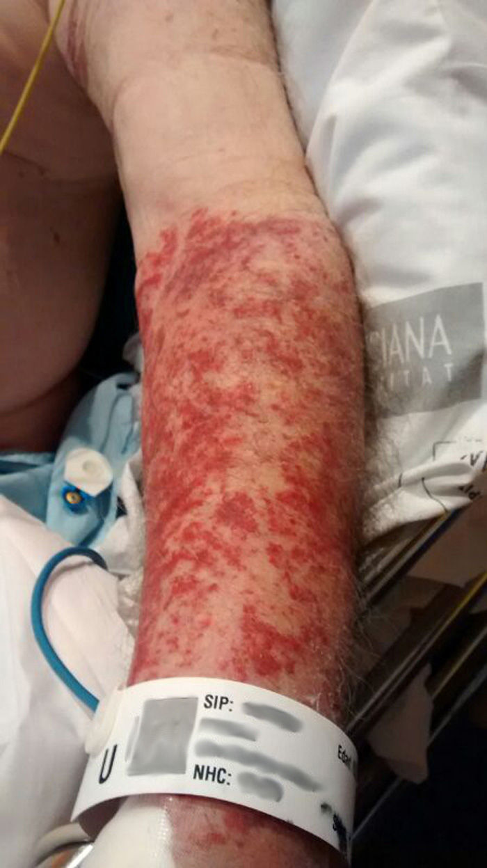
Throughout the patient's 10-h stay, his vital signs were monitored every 10min. At one point, lesions delimited by the blood pressure cuff were observed on his left arm (Fig. 1), and we decided to study their progress. Once the patient had been stabilized, he was transferred to the referral hospital for nephrological evaluation and possible dialysis due to renal failure.

During the follow-up of the patient in the referral hospital, the skin lesion spread to his other 3 limbs, and a thorough study of the pathological and clinical features, the determination of the presence of eosinophils, and a biopsy led to the diagnosis of leukocytoclastic vasculitis.
DiscussionThis phenomenon suggests 2 feasible hypotheses. The first is the possible development of ecchymosis due to malpractice in blood pressure measurement, which the patient had undergone every 5min for 10h. We have found no reports in the literature dealing expressly with secondary effects of the technique for taking blood pressure. Thus, the hypothesis that the ecchymosis we describe might be a result of this measurement could not be evaluated because of the lack of studies that confirm it.
Regarding the second hypothesis, we cannot assume that serial blood pressure readings were responsible for ecchymosis as an early sign of leukocytoclastic vasculitis without other studies that confirm the existence of this possibility. However, our experience suggests that venous stasis may have facilitated and accelerated the reactions mediated by the immune system, which responded to the allergen, in this case, the antibiotic prescribed for cystitis.
In addition to histopathological confirmation, the diagnosis of this type of vasculitis requires complete analyses of blood and urine to rule out organ involvement. This case, which was caused by a drug reaction, had an excellent prognosis. However, vasculitides associated with severe diseases have a more aggressive clinical progression, with hemorrhagic lesions, blistering and ulceration.9 The differential diagnosis should involve thrombocytopenia (although generally, in these cases, the purpuric lesions are not palpable), disseminated intravascular coagulation (more or less extensive purpura, which may or may not be palpable), scurvy (hemorrhagic follicular papules on lower extremities) and other similar purpuric dermatoses (purpuric maculae).9
We propose the need for a study of possible secondary effects of arterial blood pressure measurement; should it be found that there are really no such effects, we wonder whether this procedure might be useful for other purposes, such as the early detection of problems like vasculitis, and all those presenting with purpura. If these lesions had been considered a symptom of vasculitis, we would have been able to provide early treatment and resolution of the condition, sparing the patient from undergoing such exhaustive testing and saving on health costs. Moreover, although on this occasion, the vasculitis was not severe, had it been so, we would have been able to treat the patient promptly to ensure a good prognosis.
ConclusionGiven that routine practice rules out ecchymosis as a secondary effect of blood pressure measurement and, in view of the failure to find scientific evidence that confirms any such relationship, we suggest that ecchymosis secondary to venous stasis could be considered a warning to alert to a possible diagnosis of vasculitis. Being that vasculitis is an inflammatory process, and assuming the inflammatory theory regarding the accumulation of different immune complexes due to edema and flush, if the latter translates into a rise in pressure on the local level, and this pressure increases with the measurement of the vital sign, the consequence could be a rapid change in the clinical signs on the local level, as was observed in the case we report here. The measurement of the blood pressure every 10min over a several hours, starting soon after admission of the patient, could even be proposed as a primary screening tool to confirm or refute the differential diagnosis of vasculitis when more thorough studies have supported it, as it is not invasive and has no secondary effects.
Ethical DisclosuresProtection of human and animal subjectsThe authors declare that no experiments were performed on humans or animals for this study.
Confidentiality of dataThe authors declare that they have followed the protocols of their work center on the publication of patient data.
Right to privacy and informed consentThe authors declare that no patient data appear in this article.
Conflicts of InterestThe authors declare they have no conflicts of interest.
Please cite this article as: Ventura-Ribes O, Machancoses FH, Rosel Remírez JF. Vasculitis tras la monitorización de presión arterial. Reumatol Clin. 2016;12:216–218.


