Lucio’s phenomenon is a rare necrotizing skin reaction, almost exclusive to diffuse lepromatous leprosy, that can mimic systemic vasculitis and lead to diagnostic and therapeutic errors. We report the case of a 32-year-old woman from Paraguay with prolonged fever, hepatomegaly, and violaceous, painless necrotic skin lesions on her limbs. She was initially treated with corticosteroids for presumed vasculitis without clinical improvement. Physical examination revealed madarosis, lepromas, and altered thermal-pain sensitivity. Skin biopsy showed a perivascular histiocytic infiltrate with acid-fast bacilli, confirming multibacillary lepromatous leprosy. Treatment with rifampicin, dapsone, and clofazimine was initiated, resulting in good clinical evolution. Lucio’s phenomenon is an immune-mediated vasculopathy that may produce false-positive serologies and mimic autoimmune diseases. Histopathological confirmation is essential to avoid unnecessary immunosuppressive therapy. It should be considered in the differential diagnosis of necrotic skin ulcers, especially in patients from endemic regions. Early recognition and specific treatment help prevent severe complications.
El fenómeno de Lucio es una reacción cutánea necrotizante poco frecuente, casi exclusiva de la lepra lepromatosa difusa, que puede simular una vasculitis sistémica y conducir a errores diagnósticos y terapéuticos. Se presenta el caso de una mujer de 32 años, oriunda de Paraguay, con fiebre prolongada, hepatomegalia y lesiones cutáneas violáceas, indoloras y necróticas en extremidades. Fue tratada inicialmente como vasculitis con corticoides sin respuesta clínica. Al examen físico presentaba madarosis, lepromas y alteración de la sensibilidad termoalgésica. La biopsia cutánea mostró infiltrado histiocitario perivascular con bacilos ácido-alcohol resistentes, confirmando lepra lepromatosa multibacilar. Se instauró tratamiento con rifampicina, dapsona y clofazimina, con buena evolución. El fenómeno de Lucio es una vasculopatía inmunomediada que puede generar serologías falsamente positivas y simular enfermedades autoinmunes. La confirmación histopatológica es fundamental para evitar terapias inmunosupresoras innecesarias. Debe considerarse en el diagnóstico diferencial de úlceras cutáneas necróticas, especialmente en pacientes de áreas endémicas. El reconocimiento precoz y el tratamiento específico previenen complicaciones graves.
Lepromatous leprosy is a chronic granulomatous infection caused by Mycobacterium leprae, an obligate intracellular, acid-fast bacillus with slow replication (doubling time of 12–14 days). Transmission occurs primarily through prolonged contact with upper respiratory secretions from infected individuals, with an average incubation period of five years.1,2
The Pan American Health Organisation (PAHO) reported that, in 2023, the global number of new cases reached 182,815, with 13.6% of these reported in the Americas (24,773 new cases), where Brazil accounts for more than 90%.3
Argentina continues to be one of the countries with a high incidence of leprosy, and it is important to note that, over the last decade, prevalence and detection rates have remained almost constant. In 2023, the detection rate per 100,000 inhabitants and the prevalence rate per 10,000 inhabitants, according to the reporting jurisdiction, were .29 and .06, respectively.
The endemic areas are the North-western, North-eastern, and central provinces of the country. Although the disease is under control in Argentina, it has not been eradicated, and according to official data from the National Ministry of Health, an average of 300–400 new cases are still registered annually.4
Among the clinical presentations is the Lucio phenomenon, a serious complication observed almost exclusively in patients with diffuse non-nodular lepromatous leprosy. It is a vascular necrotising reaction mediated by immune complexes and, in some cases, by direct vascular invasion by M. leprae, leading to endothelial proliferation, vascular occlusion, and skin necrosis. Clinically, it manifests as erythematous-violaceous macules that evolve into well-defined necrotic ulcers, frequently with hypoesthesia, and can mimic cutaneous vasculitis.5–8
The aim of this publication is to present the clinical case of a patient with a lepromatous reaction (Lucio phenomenon), a rare condition in our setting, and the differential diagnoses it raises due to its resemblance to cutaneous vasculitis. The importance of making this differentiation lies not only in avoiding misdiagnosis but also in preventing the potential administration of immunosuppressants without clinical benefit and which may even worsen the infection.9,10
Clinical case reportA 32-year-old female patient from Paraguay, with no relevant medical history, was admitted to our hospital for prolonged fever, hepatomegaly, and skin lesions. The lesions were purplish and painless, some with necrotic centres, and located on her lower extremities. She had previously been treated at another institution with methylprednisolone pulses and oral corticosteroids for a presumptive diagnosis of vasculitis, without clinical response.
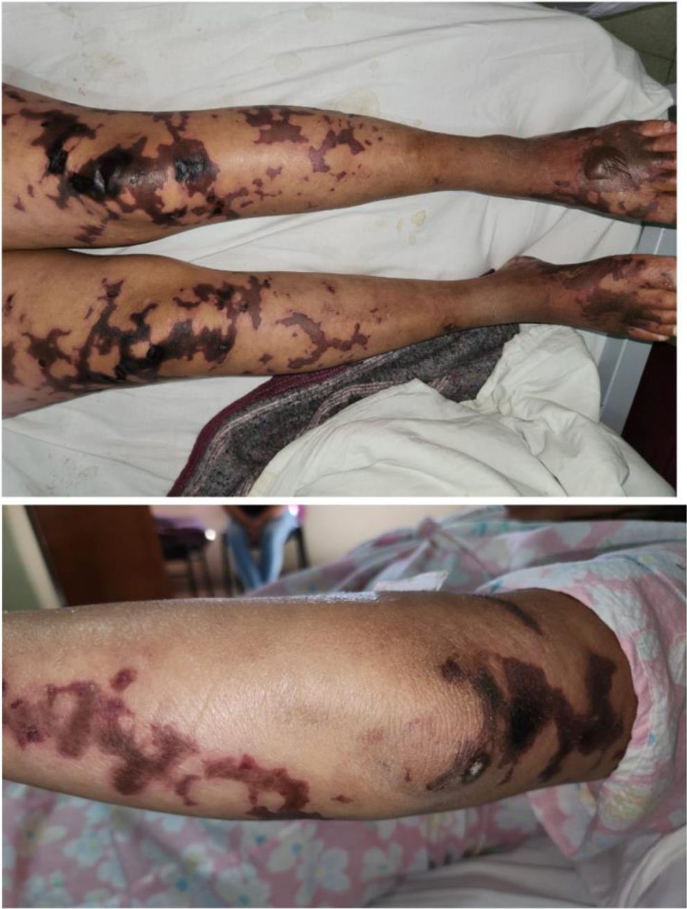
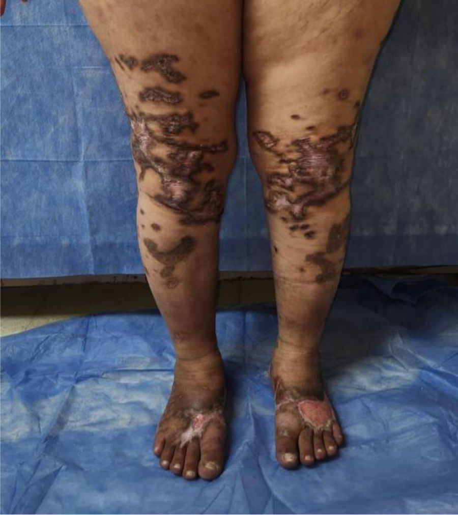
Physical examination revealed bilateral loss of the outer third of the eyebrows (madarosis), well-defined, painless, fibrinous ulcerated lesions on the earlobes and extremities, associated with hypoesthesia. A maculopapular and nodular rash (lepromas) was also observed on her face and limbs (Figs. 1 and 2). The patient reported hypoesthesia in her extremities and was therefore evaluated by the neurology service, which confirmed impaired pain and temperature sensation.


Laboratory studies revealed a ketocrit of 35.4%, hemoglobin of 10.8 g/dL, white blood cell count of 11,900/mm³, and platelet count of 350,000/mm³. Renal and hepatic function tests, urinalysis, and 24-h urine collection were unremarkable.
Serological testing was ordered for hepatitis B surface antigen (HBsAg), hepatitis B core antigen (anti-core), hepatitis C virus (HCV), and parvovirus B19 immunoglobulin M (IgM), all of which were negative. They tested positive for cytomegalovirus (CMV) and Epstein-Barr virus (EBV) immunoglobulin G (IgG). Additionally, the Venereal Disease Research Laboratory (VDRL) test was positive at the +1 dilution, and the Fluorescent Treponemal Antibody Absorption (FTA-Abs) test was positive. The purified protein derivative (PPD) was 0 mm.
Regarding the immunological analysis, the rheumatoid factor (RF) was <20, the antinuclear antibody (ANA) was 1/160 with a speckled pattern (AC-4), the complement fraction C3 was 177 mg/dL, C4 was 42 mg/dL, antineutrophil cytoplasmic antibodies (ANCA) P, C, anti-MPO, and anti-PR3 were negative, and cryoglobulins were negative.
The thrombophilia study yielded the following positive results: lupus inhibitor (LI) positive, anticardiolipin antibody (aCL) IgM >97.5 and IgG 9.2 MPL units, and anti-beta-two-glycoprotein-one antibody (aB2GP1) IgM >150 and IgG 5.4 MPL units.
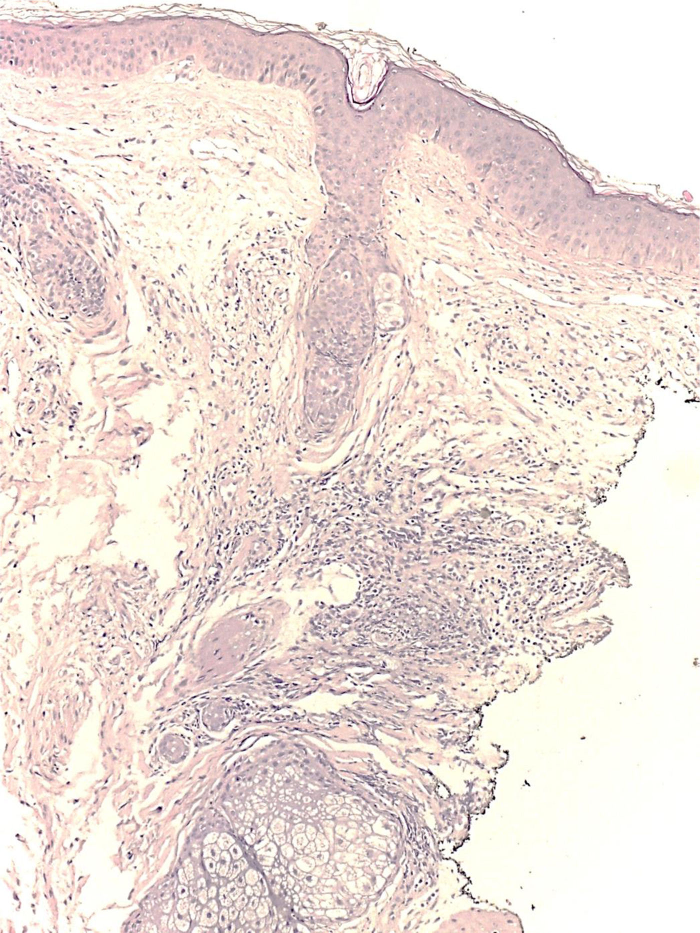
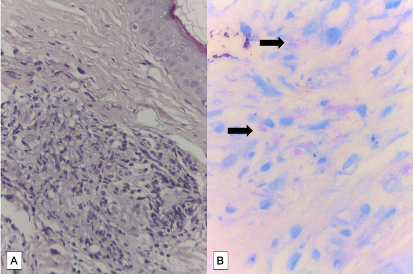
The patient was evaluated by the Dermatology Department, which performed a punch biopsy of the malar region over the nodular lesion (leproma) and of the lower limb over the ulcerated area. The histopathological report revealed chronic inflammatory infiltrate with a predominance of histiocytosis in the mid and deep dermis, with a perivascular and periadnexal distribution. Ziehl-Neelsen and Kinyoun stains were positive for acid-fast bacilli (AFB), findings consistent with lepromatous leprosy (Figs. 3 and 4). Furthermore, bacilloscopy with bacteriological and morphological index was performed, yielding positive results and confirming the diagnosis of multibacillary leprosy.


Treatment was initiated with monthly doses of rifampin 600 mg, dapsone 100 mg, and clofazimine 300 mg, followed by daily doses of dapsone 100 mg and clofazimine 50 mg for 12 months. Treatment for Lucio’s phenomenon began with meprednisone 40 mg/day in a tapering regimen. Eight months into treatment, the patient developed new nodular lesions on her face, associated with fever and malaise. This was interpreted as a type 1 reactive episode and resolved with the administration of oral corticosteroids at 40 mg/day. Twelve months into specific leprosy treatment, following the World Health Organization (WHO) regimen, she tested positive for sputum smear microscopy. Therefore, she was switched to an alternative regimen based on WHO guidelines (clofazimine, minocycline, and ofloxacin), which she continues to receive.
DiscussionLucio's phenomenon is a rare skin reaction associated almost exclusively with diffuse lepromatous leprosy (DLL), a rare variant of Hansen’s disease. It is characterised by an immune-mediated vasculopathy in which vascular occlusion secondary to the deposition of immune complexes produces dermal ischaemia and tissue necrosis. Direct vascular invasion by Mycobacterium leprae has also been documented, with endothelial proliferation and narrowing of the vascular lumen, contributing to ischaemic skin damage.5–7
Clinically, it presents as a dermatosis with abrupt onset, characterised by painful erythematous macules that rapidly evolve into purpuric lesions with geometric shapes and a livedoid pattern. These lesions may ulcerate and become necrotic, leaving atrophic, stellate scars. They typically initially affect the lower extremities and then spread in an ascending pattern. Systemic manifestations such as fever, hepatosplenomegaly, lymphadenopathy, and abnormal laboratory results may also be present, including anaemia, elevated acute-phase reactants, leukocytosis with neutrophilia, hypoalbuminaemia, hypocomplementaemia, positive rheumatoid factor, antiphospholipid antibodies, and false-positive serologies for VDRL and FTA-ABS.1,6,9
In our case, the patient presented with necrotic skin lesions on all four extremities, ear lesions, lateral madarosis, hypoesthesia, prolonged fever, and hepatosplenomegaly. Laboratory tests revealed anaemia, leukocytosis, a positive VDRL, and low titres of ANA. These findings initially led to a presumptive diagnosis of vasculitis or autoimmune disease, delaying the confirmation of diffuse lepromatous leprosy with Lucio phenomenon.
Furthermore, the antiphospholipid antibody positivity observed in our patient can be attributed to chronic infection with the bacillus. It has been reported that a large percentage of patients with leprosy, especially in its lepromatous form, present autoantibodies such as ANA and aFL, in the context of polyclonal activation of B lymphocytes and cross-reactivity to mycobacterial antigens, which are interpreted as false-positive serological findings. This finding contributes to diagnostic confusion with systemic autoimmune diseases such as systemic lupus erythematosus or antiphospholipid syndrome.10
Rodríguez-Cerdeira et al.6 described a series of 14 cases, all with severe skin lesions predominantly on the lower extremities, whose diagnosis was delayed due to their resemblance to other vasculitides. Similarly, Miranda et al.9 reported cases initially interpreted as systemic vasculitis, highlighting the importance of skin biopsy as an essential diagnostic tool.
Clinical evolution depends largely on the early initiation of specific treatment. The WHO-recommended multibacillary regimen includes rifampin 600 mg monthly and clofazimine 300 mg monthly, with daily treatment of dapsone 100 mg and clofazimine 50 mg, for 12 months.2 In Lucio phenomenon, prednisone (0.5–1 mg/kg/day) with gradual tapering is recommended, in addition to appropriate local wound care, considering that superinfection and sepsis are the main complications.5–8
Unlike other leprosy reactions, Lucio phenomenon does not always require intensive immunosuppression, which reinforces the need to differentiate it from other causes of necrotic skin ulcers, such as systemic vasculitis, cryoglobulinaemia, ulcerative infections, and neutrophilic dermatoses.
From a rheumatological viewpoint, this case highlights the importance of considering chronic infectious aetiologies as mimicking vasculitis, particularly in patients from endemic areas. Skin biopsy and histopathological, clinical, and epidemiological correlation are essential for establishing the diagnosis and avoiding unnecessary immunosuppressive therapies.
In this regard, the Ridley and Jopling classification is based on clinical and histopathological findings that correlate with the patient's immune response and the disease presentation. At one end is the tuberculoid form (preserved cellular immunity and low bacterial load), and at the other, the lepromatous form (deficient immune response, high bacterial load), with intermediate forms between these two poles (borderline). For therapeutic purposes, the WHO distinguishes between paucibacillary leprosy (smear count <2+ or up to 5 lesions and/or one affected nerve) and multibacillary leprosy (smear count ≥2+ or more than 5 lesions and/or more than one affected nerve).2–11
ConclusionLucio’s phenomenon should be considered in the differential diagnosis of necrotic skin ulcers, especially in patients from endemic areas or with compatible epidemiological backgrounds. Skin biopsy and clinical pathological correlation are essential for an accurate diagnosis. Early recognition and immediate initiation of specific treatment can prevent serious complications and neurological sequelae.
Ethical aspectsThis study adheres to the ethical principles of the Declaration of Helsinki.
The authors have no conflict of interests to declare