

The impact of smoking on osteoarthritis (OA) remains controversial. This study aimed to evaluate the effect of smoking, adjusted for body mass index (BMI) and medication use, on systemic and joint inflammation in patients with knee OA. Peripheral blood (PB), synovial fluid (SF), and histological samples were analyzed from patients stratified by smoking status, pain level (VAS), tobacco index (TI), and use of DMARDs, NSAIDs, and analgesics. IL-1β, IL-6, and TNF-α levels were measured by ELISA. Histological evaluation of extracellular matrix (ECM) used Alcian blue and Safranin O staining. Statistical analyses included ANOVA, Dunn's test, Spearman's and Kendall's correlations, and linear regression adjusted for age and BMI.
ResultsSmoking OA patients (OA-S) showed significantly higher plasma TNF-α levels compared to non-smokers (OA-NS) (P<0.01), and reduced proteoglycan content (47.15%) compared to former smokers (OA-ExS) (72.74%) (P<0.01). A moderate negative correlation was observed between SF IL-1β and proteoglycans in OA-ExS. In OA-S, plasma TNF-α strongly correlated with SF IL-6 (P<0.05). Plasma TNF-α correlated with pain in OA-S, while IL-1β did so in OA-ExS (P<0.01). BMI positively correlated with SF IL-6 in OA-S (P<0.01). Analgesic and NSAID use were associated with increased plasma TNF-α in OA-S and OA-ExS. Regression models showed that smoking was significantly associated with elevated plasma TNF-α and SF IL-6 levels, independent of BMI and age.
ConclusionSmoking, combined with excess weight and medication use, may enhance chronic inflammation and contribute to increased severity of knee OA.
El impacto del tabaquismo en la osteoartritis (OA) sigue siendo controvertido. Este estudio tuvo como objetivo evaluar el efecto del tabaquismo, ajustado por índice de masa corporal (IMC) y uso de medicamentos, sobre la inflamación sistémica y articular en pacientes con OA de rodilla. Se analizaron muestras de sangre periférica (SP), líquido sinovial (LS) e histológicas de pacientes estratificados según su condición de fumador, nivel de dolor (EVA), índice de tabaquismo (IT) y uso de FAME, AINE y analgésicos. Los niveles de IL-1β, IL-6 y TNF-α se midieron mediante ELISA. La evaluación histológica de la matriz extracelular (MEC) se realizó mediante tinción con azul de alcian y safranina O. Los análisis estadísticos incluyeron ANOVA, prueba de Dunn, correlaciones de Spearman y Kendall, y regresión lineal ajustada por edad e IMC.
ResultadosLos pacientes con osteoartritis fumadores (OA-F) mostraron niveles plasmáticos de TNF-α significativamente más altos que los no fumadores (OA-NF) (p<0,01) y un contenido reducido de proteoglicanos (47,15%) en comparación con los exfumadores (OA-ExF) (72,74%) (p <0,01). Se observó una correlación negativa moderada entre la IL-1β en líquido sinovial y los proteoglicanos en OA-ExF. En OA-F, el TNF-α plasmático se correlacionó fuertemente con la IL-6 en líquido sinovial (p <0,05). El TNF-α plasmático se correlacionó con el dolor en OA-F, mientras que la IL-1β lo hizo en OA-ExF (p <0,01). El IMC se correlacionó positivamente con la IL-6 en líquido sinovial en OA-F (p <0,01). El uso de analgésicos y AINE se asoció con un aumento del TNF-α plasmático tanto en OA-F como en OA-ExF. Los modelos de regresión mostraron que el tabaquismo se asoció significativamente con niveles elevados de TNF-α en plasma y de IL-6 en líquido sinovial, independientemente del IMC y la edad.
ConclusiónEl tabaquismo, combinado con el exceso de peso y el uso de medicamentos, puede exacerbar la inflamación crónica y contribuir a una mayor gravedad de la osteoartritis de rodilla.
Osteoarthritis (OA) is defined as a chronic disease that primarily affects individuals over 60 years old. It is considered the most prevalent joint disease characterized by cartilage volume loss, and according to the Global Burden of Disease Study (GBD), there were 303.1 million cases of hip and knee OA worldwide.1,2 This disease has been associated with several risk factors, such as overweight, sex, genetic factors, previous joint injuries, and even environmental factors like smoking.3 The role of smoking is controversial, as some observational studies have shown an inverse correlation with the development of the disease.4–6 This association may be influenced by body weight, as smoking often leads to weight loss.4–6 In contrast, other studies highlight that active smoker with OA have higher pain scores on the visual analog scale (VAS) both at baseline and during follow-up.7–9 Recently, we observed that smokers under 60 years old present severe OA, which may be mediated by an increase in arginase enzyme activity when consuming more than 10 packs per year. This was associated with a higher degree of pain and lower activity of the antioxidant enzyme glutathione-S-transferase (GST), promoting oxidative stress (OS), which is linked to the activity of matrix metalloproteinases (MMPs).10
Recent studies in human chondrocyte cultures (both monolayer and 3D) exposed to tobacco smoke extract have shown that at concentrations equivalent to smoking 10 or 20cigarettes per day, there is a significant reduction in proteoglycan content, accompanied by an increase in the expression of MMP9 and MMP13, likely mediated by IL-1β.11,12 A study by Zhang et al.,13 found an association between smoking in patients with early OA and increased serum levels of high-sensitivity C-reactive protein (hsCRP), along with the activation of pro-inflammatory cytokines, which may affect cartilage volume. This association is important since active smokers have been reported to show higher levels of IL-1β and IL-6 in plasma.14 Recently, OA has been closely linked to chronic systemic inflammation, both in early and advanced stages, contributing to pain and progression of cartilage degeneration.15,16 Additionally, smoking has been reported to negatively impact treatment responses in patients with chronic inflammatory diseases.17 This effect may be due to smoking influencing TNF-α signaling, which induces rapid gene transcription regulating inflammation via NF-kB, leading to a poor treatment response and consequently more inflammation, damage, and pain.17
However, there are still unmet needs in the field of environmental factors in OA development, highlighting the importance of conducting more studies to evaluate the effect of smoking as a risk factor and its influence on the inflammatory response in patients undergoing treatment. Therefore, the objective of this study was to determine the impact of smoking on the inflammatory process, possible cartilage deterioration, and pain levels, adjusting for the potential confounding effect of BMI and medication use in the treatment of the disease.
Materials and methodsStudy population descriptionPeripheral blood (PB) and synovial fluid (SF) samples were obtained from 51 patients with knee osteoarthritis (OA), who were classified according to smoking status: 21 non-smokers (OA-NS), 19 former smokers (OA-ExS), and 11 smokers (OA-S).
OA diagnosis was established according to the American College of Rheumatology criteria, which include age over 50 years, no morning stiffness or morning stiffness lasting less than 30min, knee crepitus, joint tenderness, osteophyte formation, and no increased temperature in the affected joint.18 Radiologically, patients were classified according to the Kellgren–Lawrence (K–L) scale,19 and pain intensity was assessed using the visual analog scale (VAS). Inclusion criteria were grades III and IV according to the K–L scale. This study was conducted in accordance with the criteria established in the Declaration of Helsinki20 and derived from the protocol, which was approved by the Ethics and Research Committee. At the time of recruitment, participants signed an informed consent form.
PB samples were collected prior to joint replacement surgery in Vacutainer tubes with EDTA-K2, and then centrifuged to separate the plasma fraction in order to obtain an accurate reading of the surrounding cytokines, which were frozen at −80°C until cytokine quantification. For SF, arthrocentesis was performed during total knee arthroplasty using syringes without anticoagulant, and aliquots were then frozen at −80°C until use. Cartilage remnants were obtained during surgery under sterile conditions and transported in phosphate-buffered saline (PBS) supplemented with 10% penicillin/streptomycin (v/v). Aliquots were made and stored at −80°C until use.
Tobacco index analysis (TI)The tobacco index (TI) was determined through a questionnaire included as part of the informed consent form. This questionnaire assessed smoking habits, including whether the individual smoked or not, as well as the quantity and duration of smoking, to calculate the tobacco index (TI). The TI was calculated by multiplying the number of cigarettes smoked per day by the number of years smoked, and dividing the result by twenty (the number of cigarettes in a pack). The number of packs consumed per year was determined, with a range categorized as greater than, less than, or equal to 10, in order to establish the effect of smoking.10
Determination of pro-inflammatory cytokinesThe quantification of IL-1β, IL-6, and TNF-α was performed using 100μl of plasma and SF samples, employing the Human Standard ABTS ELISA Development Kit (Peprotech, USA) specific for each cytokine, following the manufacturer's instructions. This included the specific capture antibody for each cytokine, detection antibodies, as well as avidin-peroxidase and the chromogenic substrate for avidin-peroxidase, 2,2′-Azino-bis (3-ethylbenzothiazoline-6-sulfonic acid) ABTS (Sigma–Aldrich, USA). The detection limits for IL-1β were 8–1000pg/ml, for IL-6 were 16–2000pg/ml, and for TNF-α were 16–2000pg/ml. Results were referenced to standard curves for IL-1β, IL-6, and TNF-α, respectively, and were expressed in pg/ml. Absorbance readings were taken using a microplate reader (Bio-Rad jMark 10176 Laboratories, Inc., USA) at 415nm.
Histological Analysis. Cartilage remnants were fixed in 4% paraformaldehyde (PFA) for 12h, dehydrated in graded ethyl alcohols (70%, 96%, and 100%), embedded in paraffin, and sectioned into 5μm thick slices using high-profile 818 blades (Leica). The sections were deparaffinized in xylene, rehydrated in a graded series of ethanol concentrations, and stained with Alcian blue (cat. 15432949) at pH 2.5 for 30min, followed by removal of excess stain with 3% acetic acid (cat. 1.00063) for 3min. The sections were counterstained with eosin (cat. 109844) for 5min to detect proteoglycans. Glycosaminoglycans were detected by staining with Safranin O (Cat. S2255) for 5min, followed by counterstaining with Fast Green (Cat. F7252) at 0.1% for 3min, and the nuclei were stained with Weigert's Hematoxylin (Cat. 115973) for 7min.12,21 Densitometric analysis was performed using ImageJ software version 1.54f (National Institutes of Health, USA) to analyze the staining intensity.22,23 The presence of proteoglycans was represented as the percentage of intensity positive for Alcian blue staining.12
Statistical Analysis. The results were analyzed using STATA version 14 (Stata Corp., College Station, TX, USA) and GraphPad version 6.0. Normality was determined using the Shapiro–Wilk test. For variables with a normal distribution, the mean and standard deviation (SD) were calculated, and for variables with an asymmetric distribution, the median and interquartile range (IQR) were calculated. The difference in the distribution of normal variables stratified by smoking habits between study groups was evaluated using analysis of variance (ANOVA) for normally distributed variables and the Dunn test for variables with an asymmetric distribution. Differences between study groups for histological sections were evaluated after stratification by smoking habit using the Kruskal–Wallis test with adjustments for multiple comparisons using the Dunn test. Correlations were assessed using the Spearman correlation coefficient adjusted by Bonferroni and the Kendall Tau coefficient for ordinal variables. To determine the association between cytokine levels and smoking habits in OA patients, multivariate linear regression models were developed, adjusting for age and BMI. A 95% confidence level was established for all analyses, and p values<0.05 were considered significant.
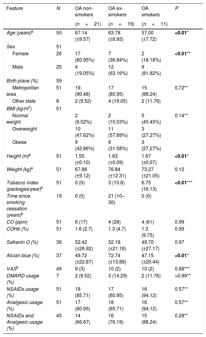
ResultsStudy population characteristicsRegarding self-reported smoking habits, three groups were established: 21 OA-NS, 19 OA-ExS, and 11 OA-S. A significant difference was observed between these groups in terms of mean age, with OA-S patients having an average age of 57±7.72 years, compared to OA-NS and OA-ExS patients who were 67.1±9.57 years and 63.78±6.92 years old, respectively (P<0.01). The proportion of male patients was 81.8% in the OA-S group, while 80.9% of the OA-NS group were female (P<0.01). Regarding BMI, 45.4% of smokers and 9.5% of non-smokers had normal weight (P=0.14). However, in terms of weight, the average for smokers was 73.3±21.05kg, in contrast to the non-smokers, who had an average weight of 67.9±9.12kg (P=0.12). Regarding the tobacco index (TI), the groups showed a significant difference in the medians, with OA-S consuming 6.75 packs per year, while OA-NS and OA-ExS consumed 0 and 3 packs annually, respectively (P<0.01). As for medication use, 86.24% of patients were under treatment with non-steroidal anti-inflammatory drugs (NSAIDs) or analgesics, while only 11.76% were taking disease-modifying anti-rheumatic drugs (DMARDs) (Table 1).
Study populations’ characteristics.
| Feature | N | OA non-smokers | OA ex-smokers | OA smokers | P |
|---|---|---|---|---|---|
| (n=21) | (n=19) | (n=11) | |||
| Age (years)a | 50 | 67.14 (±9.57) | 63.78 (±6.92) | 57.00 (±7.72) | <0.01* |
| Sex | 51 | ||||
| Female | 26 | 17 (80.95%) | 7 (36.84%) | 2 (18.18%) | <0.01** |
| Male | 25 | 4 (19.05%) | 12 (63.16%) | 9 (81.82%) | |
| Birth place (%) | 59 | ||||
| Metropolitan area | 51 | 19 (90.48) | 17 (80.95) | 15 (88.24) | 0.72** |
| Other state | 8 | 2 (9.52) | 4 (19.05) | 2 (11.76) | |
| BMI (kg/m2) | 51 | ||||
| Normal weight | 2 (9.52%) | 2 (10.53%) | 5 (45.45%) | 0.14** | |
| Overweight | 10 (47.62%) | 11 (57.89%) | 3 (27.27%) | ||
| Obese | 9 (42.86%) | 6 (31.58%) | 3 (27.27%) | ||
| Height (m)a | 51 | 1.55 (±0.10) | 1.63 (±0.09) | 1.67 (±0.07) | <0.01* |
| Weight (kg)a | 51 | 67.86 (±9.12) | 76.84 (±12.31) | 73.27 (±21.05) | 0.12 |
| Tobacco index (packages/year)b | 51 | 0 (0) | 3 (10.6) | 6.75 (16.13) | <0.01*** |
| Time since smoking cessation (years)b | 19 | 0 (0) | 21 (10–30) | 0 (0) | |
| CO (ppm) | 51 | 6 (17) | 4 (28) | 4 (61) | 0.99 |
| COHb (%) | 51 | 1.6 (2.7) | 1.3 (4.7) | 1.3 (9.75) | 0.99 |
| Safranin O (%) | 36 | 52.42 (±26.92) | 52.18 (±21.16) | 49.70 (±27.17) | 0.97 |
| Alcian blue (%) | 37 | 49.72 (±22.87) | 72.74 (±13.88) | 47.15 (±20.44) | <0.01* |
| VASb | 49 | 9 (3) | 10 (2) | 10 (2) | 0.68*** |
| DMARD usage (%) | 7 | 2 (9.52) | 3 (14.29) | 2 (11.76) | >0.99** |
| NSAIDs usage (%) | 51 | 18 (85.71) | 17 (80.95) | 16 (94.12) | 0.57** |
| Analgesic usage (%) | 51 | 17 (80.95) | 18 (85.71) | 16 (94.12) | 0.57** |
| NSAIDs and Analgesic usage (%) | 45 | 14 (66.67) | 16 (76.19) | 15 (88.24) | 0.28** |
BMI: body mass index; VAS: visual analog scale; DMARD: disease-modifying antirheumatic drugs; NSAIDs: nonsteroidal anti-inflammatory drugs (NSAIDs). Bold values denote statistical significance.
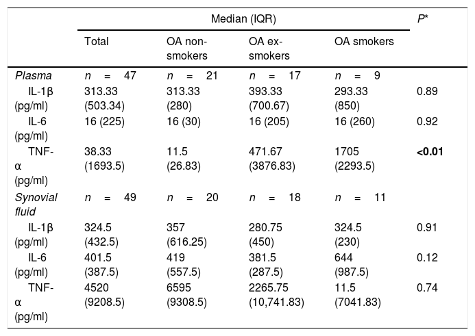
The analysis of pro-inflammatory cytokines was performed in plasma and synovial fluid (SF) according to smoking habits. A significant difference was found in the concentration of TNF-α in the OA-S group, with a value of 1705pg/ml, compared to 11.5pg/ml in the OA-NS group and 471.67pg/ml in the OA-ExS group (P<0.01) at the systemic level. However, in the SF analysis, only IL-6 showed a slight increase in the OA-S group, with a concentration of 644pg/ml, compared to 419pg/ml in OA-NS and 381.5pg/ml in OA-ExS. Nevertheless, this difference was not statistically significant (P=0.12) (Table 2).
Cytokine concentrations by anatomical measurement site and smoking habit.
| Median (IQR) | P* | ||||
|---|---|---|---|---|---|
| Total | OA non-smokers | OA ex-smokers | OA smokers | ||
| Plasma | n=47 | n=21 | n=17 | n=9 | |
| IL-1β (pg/ml) | 313.33 (503.34) | 313.33 (280) | 393.33 (700.67) | 293.33 (850) | 0.89 |
| IL-6 (pg/ml) | 16 (225) | 16 (30) | 16 (205) | 16 (260) | 0.92 |
| TNF-α (pg/ml) | 38.33 (1693.5) | 11.5 (26.83) | 471.67 (3876.83) | 1705 (2293.5) | <0.01 |
| Synovial fluid | n=49 | n=20 | n=18 | n=11 | |
| IL-1β (pg/ml) | 324.5 (432.5) | 357 (616.25) | 280.75 (450) | 324.5 (230) | 0.91 |
| IL-6 (pg/ml) | 401.5 (387.5) | 419 (557.5) | 381.5 (287.5) | 644 (987.5) | 0.12 |
| TNF-α (pg/ml) | 4520 (9208.5) | 6595 (9308.5) | 2265.75 (10,741.83) | 11.5 (7041.83) | 0.74 |
Bold values denote statistical significance.
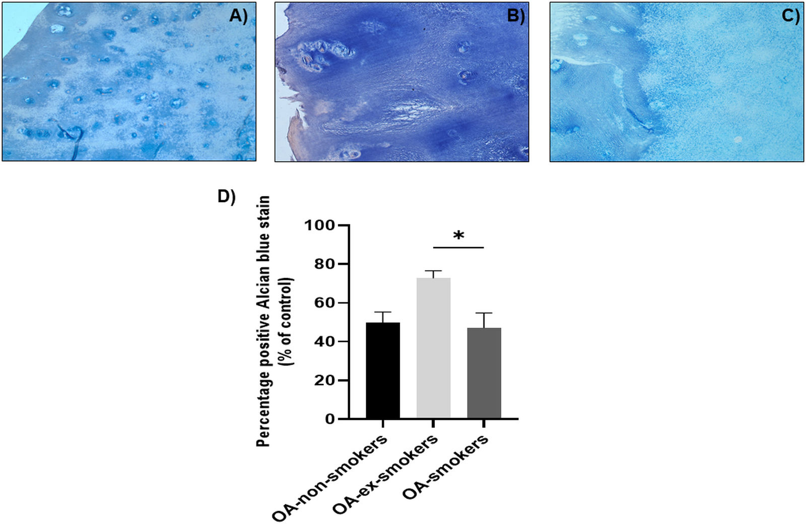
The composition of proteoglycans was evaluated using Alcian blue staining. The OA-S group showed a decrease in the intensity of Alcian blue, with a value of 47.15%, compared to 49.72% and 72.74% in the OA-NS and OA-ExS groups, respectively (P<0.01) (Table 1, Fig. 1). No significant changes were observed in the composition of glycosaminoglycans between the groups stratified by smoking habit (Table 1).
Effect of smoking on the presence of proteoglycans in the articular cartilage of patients with knee OA. Alcian blue staining evaluated according to smoking status, with panel (A) non-smoking patient, (B) former smoker, and (C) current smoker. Representative images from 37 histological sections. (D) Percentage expression of proteoglycan presence showing a significant decrease in smoking patients (47.15%) compared to former smokers (72.74%) (*P<0.01). Densitometric analysis was performed using ImageJ version 1.54f to determine the percentage of positive staining. Dunn's test.
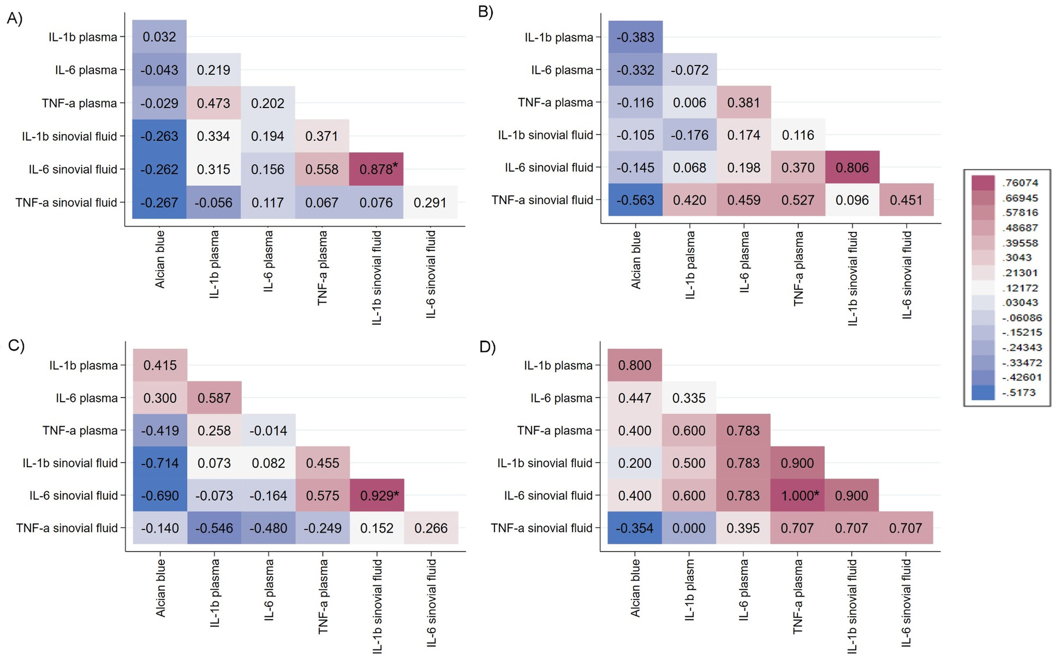
Spearman correlation coefficients were estimated to assess whether smoking affected the inflammatory response at both the plasma and joint levels, considering the synovial fluid (SF) and proteoglycans in articular cartilage. The correlation between cytokine levels in the entire study population, without stratification, showed a significant positive correlation between IL-6 and IL-1β in SF (Spearman's rho=0.878, P<0.001) (Fig. 2A). When stratified by smoking habit, the OA-ExS group showed a significant positive correlation between IL-6 and IL-1β in SF, with a rho value higher than that of the total cytokines (Spearman's rho=0.929, P=0.02). A negative correlation was also found between IL-1β and IL-6 levels and the presence of proteoglycans (Spearman's rho=−0.714 and −0.690, respectively); however, after adjusting for Bonferroni, statistical significance was not reached (P=0.72 and P=0.63, respectively) (Fig. 2C). When cytokines were compared in the OA-S group at the plasma level, TNF-α perfectly correlated with IL-6 levels (Spearman's rho=1.00, P<0.001), as well as with IL-1β, though not significantly (Spearman's rho=0.900, P=0.55) (Fig. 2D).
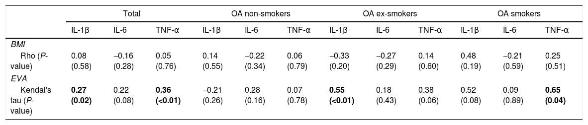
Cytokine correlation in plasma in relation to smoking habit, BMI, and VASWhen evaluating the correlation of total IL-1β and TNF-α concentrations (obtained from the sum of plasma and SF concentrations) with respect to the Visual Analog Scale (VAS), these cytokines showed a positive association with a Kendall's Tau coefficient of 0.27 (P=0.02) and 0.36 (P<0.01), respectively. Although statistically significant, the correlation was weak. Regarding the OA-S group, TNF-α concentration, with respect to VAS, showed a positive relationship with a Kendall's Tau of 0.65 (P=0.04). As for the OA-ExS group and VAS, a significant positive correlation was found, with a Kendall's Tau value of 0.55 (P<0.01) (Table 3).
Correlation between the concentration of cytokines in plasma and BMI and VAS by study group.
| Total | OA non-smokers | OA ex-smokers | OA smokers | |||||||||
|---|---|---|---|---|---|---|---|---|---|---|---|---|
| IL-1β | IL-6 | TNF-α | IL-1β | IL-6 | TNF-α | IL-1β | IL-6 | TNF-α | IL-1β | IL-6 | TNF-α | |
| BMI | ||||||||||||
| Rho (P-value) | 0.08 (0.58) | −0.16 (0.28) | 0.05 (0.76) | 0.14 (0.55) | −0.22 (0.34) | 0.06 (0.79) | −0.33 (0.20) | −0.27 (0.29) | 0.14 (0.60) | 0.48 (0.19) | −0.21 (0.59) | 0.25 (0.51) |
| EVA | ||||||||||||
| Kendal's tau (P-value) | 0.27 (0.02) | 0.22 (0.08) | 0.36 (<0.01) | −0.21 (0.26) | 0.28 (0.16) | 0.07 (0.78) | 0.55 (<0.01) | 0.18 (0.43) | 0.38 (0.06) | 0.52 (0.08) | 0.09 (0.89) | 0.65 (0.04) |
BMI: body mass index; VAS: visual analog scale. Bold values denote statistical significance.
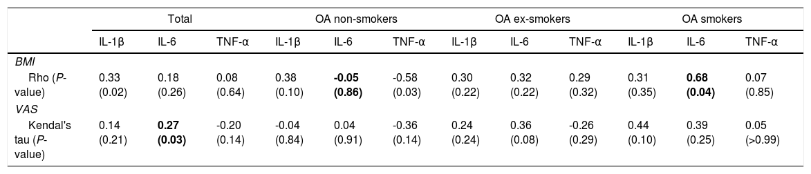
When evaluating the correlation between the total IL-6 level (obtained from the sum of IL-6 concentrations in plasma and SF) and the VAS, we found a significant positive concordance of 0.27 (P=0.03) using Kendall's Tau coefficient. Regarding BMI, the OA-S group showed a positive correlation with the IL-6 concentration in SF (Spearman's rho=0.68, P<0.04). In comparison, the OA-NS group exhibited a negative correlation between BMI and TNF-α concentration, with a Spearman's rho value of -0.58 (P<0.03) (Table 4).
Correlation between the concentration of cytokines in synovial fluid and BMI and VAS by study group.
| Total | OA non-smokers | OA ex-smokers | OA smokers | |||||||||
|---|---|---|---|---|---|---|---|---|---|---|---|---|
| IL-1β | IL-6 | TNF-α | IL-1β | IL-6 | TNF-α | IL-1β | IL-6 | TNF-α | IL-1β | IL-6 | TNF-α | |
| BMI | ||||||||||||
| Rho (P-value) | 0.33 (0.02) | 0.18 (0.26) | 0.08 (0.64) | 0.38 (0.10) | -0.05 (0.86) | -0.58 (0.03) | 0.30 (0.22) | 0.32 (0.22) | 0.29 (0.32) | 0.31 (0.35) | 0.68 (0.04) | 0.07 (0.85) |
| VAS | ||||||||||||
| Kendal's tau (P-value) | 0.14 (0.21) | 0.27 (0.03) | -0.20 (0.14) | -0.04 (0.84) | 0.04 (0.91) | -0.36 (0.14) | 0.24 (0.24) | 0.36 (0.08) | -0.26 (0.29) | 0.44 (0.10) | 0.39 (0.25) | 0.05 (>0.99) |
BMI: body mass index; VAS: visual analog scale. Bold values denote statistical significance.
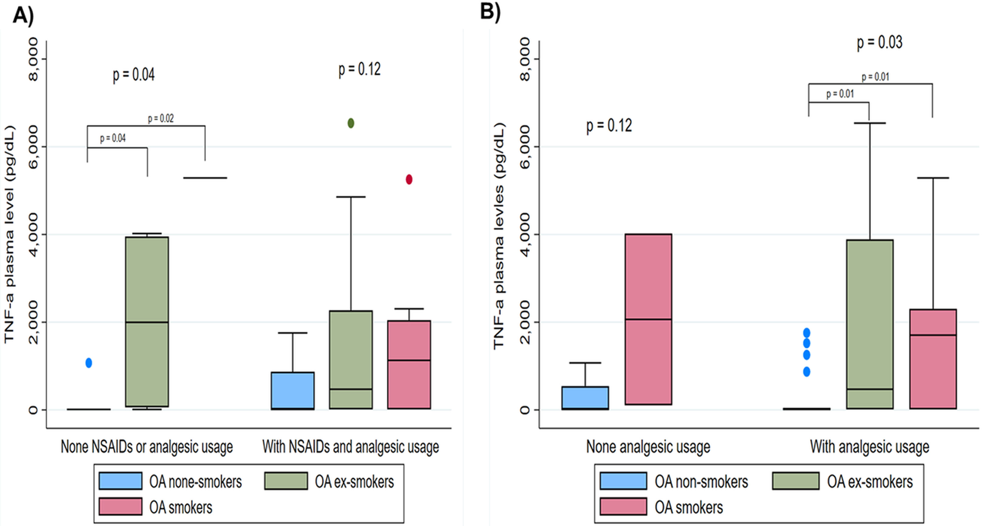
When evaluating TNF-α levels in plasma and SF in relation to treatment and smoking habits, we observed that patients who were not using NSAIDs or analgesics in the OA-ExS group had higher TNF-α levels (5288.33pg/ml) compared to the OA-NS group (11.5pg/ml) and the OA-S group (1996.665pg/ml) (P=0.04). When evaluating this cytokine among patients treated with NSAIDs and analgesics, no difference in concentration was observed when stratifying by smoking habit (P=0.12) (Fig. 3A). However, when comparing plasma TNF-α levels by smoking habit in patients taking analgesics regardless of NSAID use the OA-ExS group showed the highest concentration (p50=1705pg/ml), followed by the OA-S group (p50=471.67pg/ml), while the OA-NS group showed no TNF-α production (p50=11.5pg/ml) (Fig. 3B).
TNF-α levels associated with smoking status and treatment. (A) Total TNF-α concentration in relation to the use of NSAIDs and analgesics, or no treatment, showed a significant increase in former smokers (P=0.04). (B) Plasma TNF-α concentration showed that patients who take analgesics and have a history of smoking exhibit elevated levels compared to non-smokers (P=0.03).
When evaluating TNF-α concentration in SF, as well as IL-1β and IL-6 levels, no significant differences were observed between smoking habit groups when stratified by the use of analgesics, NSAIDs, or the combination of both.
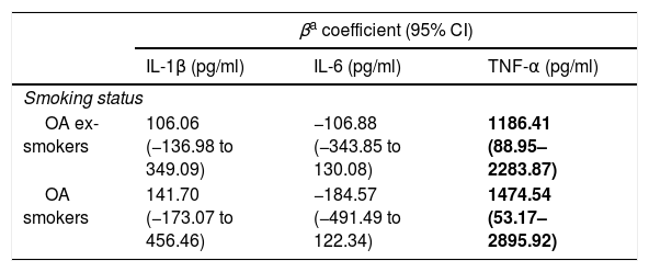
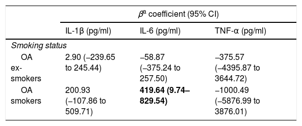
Estimated mean difference in cytokine concentration in plasma and SF according to smoking habitWhen performing linear regression models between cytokine concentrations in plasma and smoking habits adjusting for age and BMI to assess the direct relationship between smoking and the disease, we found no significant association with IL-6 or IL-1β. However, we observed that OA-S individuals had 1474.54pg/ml more TNF-α than OA-NS individuals (P=0.042), and even OA-ExS showed 1186.41pg/ml more TNF-α than OA-NS (P=0.032) (Table 5). In SF, we found that OA-S individuals had 419.64pg/ml more IL-6 than OA-NS (P=0.045) (Table 6).
Average estimated difference in the concentration of cytokines evaluated in plasma by smoking habit.
| βa coefficient (95% CI) | |||
|---|---|---|---|
| IL-1β (pg/ml) | IL-6 (pg/ml) | TNF-α (pg/ml) | |
| Smoking status | |||
| OA ex-smokers | 106.06 (−136.98 to 349.09) | −106.88 (−343.85 to 130.08) | 1186.41 (88.95–2283.87) |
| OA smokers | 141.70 (−173.07 to 456.46) | −184.57 (−491.49 to 122.34) | 1474.54 (53.17–2895.92) |
Average estimated difference in the concentration of cytokines evaluated in synovial fluid by smoking habit.
| βa coefficient (95% CI) | |||
|---|---|---|---|
| IL-1β (pg/ml) | IL-6 (pg/ml) | TNF-α (pg/ml) | |
| Smoking status | |||
| OA ex-smokers | 2.90 (−239.65 to 245.44) | −58.87 (−375.24 to 257.50) | −375.57 (−4395.87 to 3644.72) |
| OA smokers | 200.93 (−107.86 to 509.71) | 419.64 (9.74–829.54) | −1000.49 (−5876.99 to 3876.01) |
Recently, the U.S. National Health Interview Survey reported that 14 million people suffer from symptomatic knee OA. Thus, identifying potentially modifiable risk factors may lead to strategies that help delay OA progression.24
Among the modifiable factors, smoking has been a controversial one regarding OA development. Some in vitro and in vivo models, as well as clinical studies, have associated smoking with the degree of pain in OA patients.11,25 In contrast, a meta-analysis indicated a protective association between smoking and OA development, although BMI was not adjusted for.4 The lack of BMI adjustment is crucial, as smokers tend to be thinner than non-smokers, and obesity is a well-established risk factor for OA.26 On the other hand, clinical studies in various populations have shown that smoking is negatively associated with OA incidence.5,27,28 Recently, a study using multivariable Mendelian randomization, adjusted for BMI, reported that smoking has an independent harmful causal effect on OA development, highlighting the importance of smoking cessation and obesity management to reduce OA incidence.29
Our study reveals that a higher proportion of smokers are of normal weight compared to non-smokers and former smokers. Furthermore, fewer smokers were overweight relative to non-smokers and former smokers. When analyzing obesity incidence, we found a lower proportion among smokers compared to those who quit or never smoked. These results suggest that smoking affects BMI but does not appear to provide protection against OA development, as both smokers and former smokers reported higher pain levels compared to those who never smoked. This occurred even though both OA groups had the highest use of NSAIDs and analgesics 88.24% and 76.19%, respectively compared to 66.67% in patients who never smoked. These results contrast with a study from the UK that described a protective role of smoking, noting weight gain within 12 months after smoking cessation, which promoted knee cartilage degeneration.30 In our study, although the former smoker group reported more pain, they had a higher percentage of proteoglycans compared to current smokers. This might be due to a lower annual cigarette consumption (3 packs) compared to current smokers (approximately 7 packs). This aligns with a previous finding where smoking more than 10 packs increased arginase activity an enzyme considered a biomarker of disease progression and severity. Arginase has recently been reported to promote intervertebral disk degeneration via oxidative stress. However, it is important to propose an in vivo mouse model exposed to cigarette smoke to evaluate the enzyme's activity over time and correlate it with cartilage degeneration.10,31,32
A Mendelian randomization analysis supports our hypothesis, stating that the prolonged consumption of more cigarettes increases the risk of OA. A prospective cohort found that smokers had a 2.20-fold higher risk of OA compared to non-smokers.33 This finding is also supported by a cross-sectional study in the U.S., which reported a 54% higher risk in smokers and a 38% higher risk in former smokers for developing OA. This evidence supports the negative impact of smoking on OA prevalence.24,33,34
Regarding the inflammatory response, we found significantly higher plasma TNF-α concentrations in smokers compared to non-smokers and former smokers. This cytokine is a known indicator of inflammation, as it can stimulate its own production. In the joint environment, it induces chondrocytes and synoviocytes to produce other cytokines such as IL-6, contributing to cartilage degradation.35 According to Lv et al.,36 increased cytokine concentrations are associated with pain and morning stiffness. This finding is relevant, as a recent study by Skrzypkowska et al.37 reported a plasma TNF-α level of 23.8pg/ml in non-smoking OA patients. Our findings suggest that smoking may be an additional risk factor that increases pro-inflammatory cytokines like TNF-α. Even after smoking cessation, these levels may remain elevated for some time.
In SF, no significant changes were observed; however, IL-6 showed a trend toward higher concentrations in smokers. Compared to Tsuchida et al.,38 who reported IL-6 levels of 396pg/ml in non-smoking OA patients, our OA-S group had levels 248 times higher. This suggests that smoking may increase IL-6, which is known to inhibit proteoglycan synthesis and stimulate aggrecanase production.39 Nevertheless, to confirm smoking's role in elevating cytokines, a longitudinal study is needed to evaluate cytokine levels in smokers, former smokers, and non-smokers from diagnosis through follow-up. This would help assess disease development and progression related to cytokines induced by smoking.
When correlating plasma and SF cytokines, we found that in former smokers there was a significant positive correlation between IL-6 and IL-1β in the joint, along with a non-significant negative correlation between IL-1β and IL-6 in SF and proteoglycan presence. In smokers, a very strong and significant correlation was found between plasma TNF-α and SF IL-6 levels. Additionally, in SF, IL-6 was significantly associated with IL-1β. This supports the findings of Kou and Wu,35 who noted that TNF-α stimulates chondrocytes and synoviocytes to produce other cytokines, and Lv et al.,36 who consider these markers to be associated with symptomatic OA progression.
To highlight the impact of smoking on pro-inflammatory cytokine levels and their correlation with VAS and BMI, we found that systemic TNF-α was significantly correlated with pain severity in smokers. Additionally, IL-1β in former smokers showed a significant correlation. Regarding IL-6, at the SF level, there was a significant positive correlation with BMI in OA smokers, suggesting that being overweight and smoking may synergistically promote joint inflammation. While no significant correlations were found in SF, both IL-1β and IL-6 showed a positive correlation with pain, suggesting that smoking may adversely affect the joint, leading to greater pain in these patients. These findings align with those reported by Zhang et al.,13 who found that male knee OA patients who smoked experienced more intense pain.
Notably, we observed that TNF-α levels are influenced by smoking even when the patient is undergoing NSAID treatment. When comparing untreated patients, smokers showed no detectable TNF-α levels, in contrast to those taking NSAIDs. This suggests that smoking may not only be a risk or aggravating factor but may also affect anti-inflammatory drug response. However, this should be confirmed in a study that measures cytokine levels before and after treatment to assess whether smoking influences drug efficacy.
Finally, to assess the likelihood of developing OA in smokers based on cytokine levels, we found that plasma TNF-α is strongly associated with disease, while IL-6 at the local level contributes to cartilage damage potentiated by smoking. However, our findings should be interpreted cautiously due to the small sample size. Future studies will expand the study population to strengthen these preliminary results. Additionally, we will evaluate anti-inflammatory cytokines and molecules involved in the inflammatory response, such as kinase pathways and Toll-like receptors. Immunohistological analyses of metalloproteinase expression will also be conducted to assess cartilage damage exacerbated by smoking through inflammatory responses. Importantly, this preliminary study suggests that measuring cytokine levels not only in plasma but also in SF could improve or support OA prognosis in smoking patients.
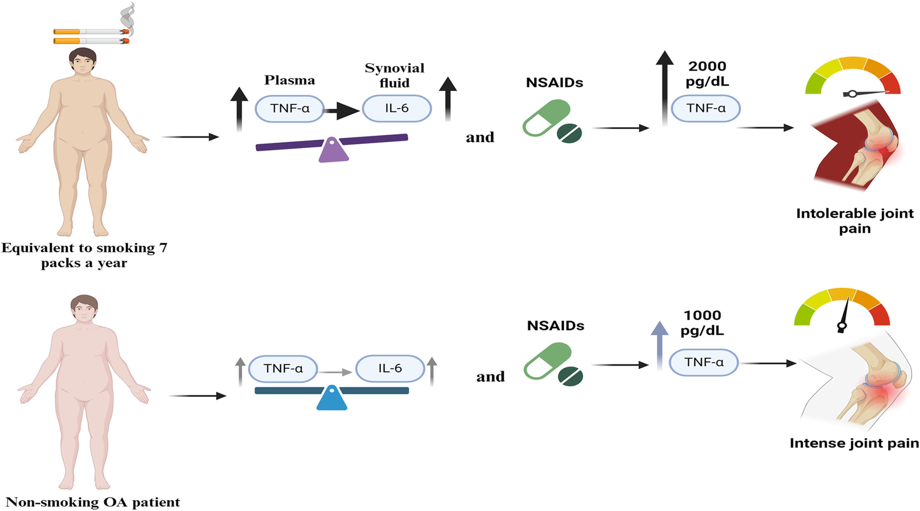
ConclusionThis study suggests that tobacco consumption has a negative impact on the inflammatory response in patients with OA, enhancing inflammation and thereby exacerbating disease symptoms. As a result, smoking patients experience a greater degree of pain associated with TNF-α, which could lead to an early decline in their quality of life. These findings highlight the importance of considering smoking as an aggravating factor in OA management, suggesting that smoking cessation could improve therapeutic outcomes and reduce pain in this patient group (Fig. 4).
Effect of smoking on the inflammatory response in patients with osteoarthritis. In patients who smoke 7 or more packs per year, there is a significant increase in plasma TNF-α, which promotes the elevation of IL-6 in the synovial fluid. This effect is further enhanced by the use of analgesics, with or without NSAIDs, reaching TNF-α concentrations up to 471.67pg/ml, associated with unbearable pain. In contrast, non-smoking patients do not show a significant increase in TNF-α and IL-6 levels, even with the use of analgesics with or without NSAIDs, showing TNF-α levels of 11.5pg/ml and experiencing intense but not unbearable pain.
JFT and GAMN: designed the study and the experimental component, analyzed, and interpreted the results to create the manuscript, and gave approval for the final version of the manuscript. AAMV and YZC: contributed to the analysis of the results at the level of cytokine expression. CGHV and CMTM: supported the analysis of IL-1β, IL-6 and TNF-α and reviewed the literature for inflammation and smoking in osteoarthritis studies. BVS, JFCM and ALM: contributed to the structuring and drafting of the manuscript, and in conducting histological techniques and analyses. VIS and REM: diagnosed OA using radiological assessments and the KL scale, supported the preparation of the samples and reviewed the literature for the discussion of the results. KMF: contributed to the study design, experiment oversight, statistical analysis for interpretation of the results, manuscript preparation and approval of the final revision of the manuscript.
Ethical approval and consent to participateAll subjects signed an informed consent before being enrolled. The study was approved by the Bioethical Committee of the National Rehabilitation Institute (protocol number 67/19).
Consent to participateInformed consent was obtained from all individual participants included in the study.
Consent for publicationAll authors have read and agreed to the published version of the manuscript.
FundingThis research did not receive any specific grant from funding agencies in the public, commercial, or not-for-profit sectors.
Conflict of interestThe authors declare that they have no financial or personal conflicts of interest in relation to this work.
We are grateful for the support in the collection of samples from EER Diana Cepeda García, Head of the Servicio de DNM e Infecciones Óseas, EER María de Jesús Hernández Rocha, Head of Enfermería de Quirófano de Cirugía Articular, and EER Miriam Rojo Maldonado and EER Gloria González Estrada, Área de Cirugía Articular. We are grateful for the assistance of Dr. Raúl Pichardo Bahena in performing histological techniques in the Anatomic Pathology and Electron Microscopy Laboratory. We are grateful to the Instituto Nacional de Rehabilitación “Luis Guillermo Ibarra Ibarra” (INRLGII).