



Minor salivary gland biopsy (MSGB) plays a pivotal role in the classification and diagnosis of Sjögren’s disease, one of the most prevalent systemic autoimmune disorders within the spectrum of rheumatic diseases. Furthermore, it provides valuable phenotypic information about the patient, contributes to the assessment of disease prognosis, and serves as a reliable tool for monitoring therapeutic response. MSGB is also useful in the diagnosis of amyloidosis, sarcoidosis, IgG4 disease, or lymphoma. In recent years, less invasive techniques have become increasingly widespread and have been proposed as highly accessible procedures for rheumatologists—straightforward to perform and associated with minimal or no complications. Consequently, MSGB has become a key diagnostic and follow-up tool in the comprehensive management of the disease.
La biopsia de glándula salival menor (BGSM) es fundamental en la clasificación y diagnóstico de la enfermedad de Sjögren (ESj), una de las enfermedades autoinmunes sistémicas más frecuentes entre las enfermedades reumáticas. Además, aporta información fenotípica del paciente, información sobre el pronóstico de la enfermedad y es una herramienta de monitorización de respuesta a tratamientos. La BGSM es también de utilidad en el diagnóstico de la amiloidosis, la sarcoidosis, la enfermedad por IgG4 o el linfoma. Las técnicas de mínima incisión han proliferado en los últimos años y se postulan como procedimientos muy accesibles para el reumatólogo, fáciles de realizar, con pocas o nulas complicaciones. Esto convierte a la BGSM en una prueba clave tanto al inicio como en el seguimiento de la enfermedad.
Minor salivary gland biopsy (MSGB) is fundamental to the classification and diagnosis of Sjögren's syndrome (SS) as it enables the detection and quantification of the inflammatory infiltrate that occurs in the exocrine glands — the target organs of this disease. SS is one of the most common and heterogeneous systemic autoimmune diseases. It is characterised by dry eyes and mouth, B-cell hyperactivity, multiple extraglandular manifestations and the development of lymphoproliferative disease.1 The first cohort of patients with SS was published in 1933 by the Swedish ophthalmologist Henrik Sjögren, after whom the disease is named.2 MSGB plays a prominent role in the classification criteria for SS, being one of the two major criteria.3 Numerous studies have highlighted the association between MSGB findings and the development of systemic involvement and lymphoma.4 MSGB provides valuable information on the patient's phenotype and prognosis, and is useful as a tool for monitoring therapeutic response. It is also the subject of study in the omics sciences.
This article aims to highlight the multiple uses of MSGB beyond diagnosing SS. A 'minimal incision' technique is described for performing MSGB in a minimally invasive manner without sutures, with few or no complications. This allows rheumatologists to perform the procedure more safely and frequently.
Histological findingsFocal lymphocytic sialadenitisThe characteristic histological pattern of Sjögren’s syndrome (SS) is focal lymphocytic sialadenitis (FLS) (Fig. 1), which is defined as an inflammatory infiltrate organised in dense aggregates in perivascular and/or periductal areas adjacent to normal acini.5,6 However, the FLS pattern can also be observed in other systemic autoimmune diseases and in healthy individuals, meaning it is not diagnostic of SS on its own. Another relatively common pattern in older individuals is chronic nonspecific sialadenitis (CNS), characterised by acinar atrophy, interstitial fibrosis, and ductal dilatation. The presence of CNS neither rules out nor confirms the presence of SS; it may represent the final stage of SS, or it may be secondary to another condition.7

Histology image of the minor labial salivary gland.
Minor salivary gland tissue represented by four acini with intense lymphocytic inflammatory infiltrate, forming numerous aggregates of more than 50 lymphocytes. These findings are consistent with focal lymphocytic sialadenitis, suggestive of Sjögren’s syndrome.
Recommendations have been described and published on the basic requirements of the biopsy sample for correct histological evaluation. These include an adequate number of lobules (four to six) and a minimum surface area of 8mm2). Specific findings in SS are also defined: a focus would be defined as an aggregate of ≥50 mononuclear cells (mainly lymphocytes)6 (Fig. 1). After identifying the pattern suggestive of SS, the number of foci per 4mm2 area or focus score (FS) can be calculated, a histological measure that quantifies glandular inflammation (FS=number of foci of ≥50 lymphocytes/area evaluated in mm2 × 4).8 There is a correlation between the different histological classification systems, such that an FS of 1 corresponds to grade 3 of the classic classification system described by Chisholm and Mason in 1968.5
Other histological lesions in SSLymphoepithelial lesions (LELs) are defined by the presence of lymphocytes in the ducts, leading to hyperplasia of the epithelium of the striated ducts in the salivary glands. They are detected using immunohistochemical techniques and are considered to be characteristic of SS; however, their sensitivity and specificity are unknown.9 Germinal centre (GC) like lesions are well-organised lymphocytic aggregates characterised by the formation of areas of active B-cell proliferation and networks of follicular dendritic cells.10 Described as characteristic of SS in 2003, their prevalence is estimated at 20%–25%.11 Immunohistochemistry techniques aid in their recognition,12 although their standardisation is still pending within the scientific community. Plasma cell displacement is defined as an increase in the number of IgG plasma cells compared to the number of IgA plasma cells, which remain stable. The diagnostic threshold for a relative decrease of <70% in IgA was established by Bodeutsch in 199213 and has high sensitivity (94.5%) and specificity (100%) for SS.14 Van Ginkel et al.15 have described the added diagnostic value of these three lesions for FS: for compliance with the 2016 classification criteria,3 specificity increased from 84% to 95% when an abnormal biopsy was defined by the presence of two or more of these four histopathological features, rather than just FS≥1.
Stratification and phenotyping of patients with SSThe clinical and serological heterogeneity of Sjögren's syndrome has driven research into phenotypic determinants that help us stratify patients. Histological characterisation of the salivary glands provides information on the severity and systemic manifestations of SS.4 An FS≥1 has been associated with reduced stimulated and unstimulated salivary flow, worsening of the Schirmer test at the five-year follow-up, and poorer oral health outcomes, (caries, tooth loss, and dental fillings). A significant correlation has been reported between a higher FS score and positivity for antinuclear antibodies, rheumatoid factor, Anti-Ro/La antibodies, hypergammaglobulinaemia, low C4 levels, cytopenias, vasculitis, Raynaud's phenomenon, lymphadenopathy, and splenomegaly. An FS≥4 has been associated with a higher prevalence of interstitial lung disease and airway involvement.16,17 An FS≥4 is a histological measure and a predictive biomarker for the early diagnosis of lymphoma in SS, as described by Chatzis et al.18
Monitoring response to treatmentMSGB performed before and after treatment in clinical trials on SS provide information on treatment efficacy, the mode of action of drugs, and the pathogenic mechanism of inflammation in glandular tissue.14 Pre-treatment histological findings can inform the personalised choice of drug. For example, Delli et al. found that a high number of CD20+/mm2 B cells in the parotid gland prior to treatment predicts a better response to rituximab.19 Standardisation of this practice is essential in future to allow adequate comparison between studies.
Other advantages of MSGBThe 'secretome', or salivary gland tissue supernatant, enables the identification of numerous inflammatory mediators that could facilitate the discovery of therapeutic targets and the development of diagnostic, prognostic, and treatment response markers.20 The omics sciences applied to glandular tissue enable the construction of cellular and spatial maps of the key components involved in the development and perpetuation of the disease, as well as the identification of molecular and functional interactions that may provide therapeutic clues.21 The labial and parotid glands in SS contain similar key histopathological features; therefore, both glands can be used for diagnosing and classifying SS.22 Furthermore, MSGB may be useful in diagnosing amyloidosis, sarcoidosis, IgG4 disease, or lymphoma, among others. All of the above has led experts to suggest that MSGB should be performed as standard practice for all patients with SS.14
Minimal incision technique: labial gland biopsyMSGB has been used as a tool for evaluating SS for more than 50 years, as described by Chisholm and Mason in 1968.5 With the patient seated in a chair and after local anaesthesia has been administered, an elliptical horizontal segment of oral mucosa containing labial salivary glands is removed.
The glands of the lower lip are easily accessible, located above the muscle layer, separated from the mucous membrane by a thin layer of fibrous connective tissue, and the possibility of excessive bleeding is minimal.23 The biopsy wound was closed with 04-gauge black silk sutures. This technique is still used today and is advocated by some authors,24,25 although several techniques have been described over the years, including small linear incisions, punch biopsy, and superficial needle biopsy,26 as well as the use of a smart lip holder for minimally invasive MSGB.27
In 2014, Varela Centelles et al. published a study comparing the prevalence of neurological complications related to lip biopsies for diagnosing SS using conventional versus minimally invasive techniques. They conducted a systematic review and meta-analysis of prevalence and concluded that the minimally invasive lip biopsy technique is associated with fewer permanent neurological complications than conventional techniques involving large linear incisions.28
Informed consentAs with any other surgical procedure, the patient must sign an informed consent form prior to the procedure. This form explains the technique and possible side effects. Patients do not need to fast before the procedure. It is recommended that the patient does not wear lipstick. It is essential to establish whether the patient is allergic to local anaesthetics, as this would contraindicate their use. It is advisable to know if the patient is taking antiplatelet or anticoagulant medication, which does not need to be discontinued and does not contraindicate the procedure. After the procedure, patients are advised not to eat solid foods or drink hot liquids for 2−3h following the intervention. They may drink cold liquids and perform normal oral hygiene, but should avoid rubbing or cleaning the wound to prevent it from opening. It is essential to record the procedure in the medical record.
Preparation of materials and spaceWith regard to space, MSGB – labial – is a low-risk, superficial, minimally invasive procedure performed under local anaesthesia, without the need for sedation, advanced monitoring or complex surgical equipment. Therefore, an operating theatre or special room is NOT required, provided that the regulatory conditions for a consultation room equipped for minor procedures are met. This type of procedure is considered minor outpatient surgery and can be performed safely in a medical consultation with the appropriate means and the capacity to manage minor complications (minor bleeding, pre-syncope/vasovagal syncope). It is advisable to have a stretcher in the room on which to lay the patient in case they feel dizzy, and adrenaline ampoules in case of a severe allergic reaction to the local anaesthetic.
For the procedure, a chair is needed in the room for the patient to sit on; behind the patient, a trained person will be positioned to hold the everted and taut lower lip in place, in our case, the rheumatology nurse (Fig. 2). It is advisable to have an auxiliary light source. The following materials should be placed on a side table: sterile gloves, sterile scalpel, sterile straight forceps without teeth (the finer they are, such as those used in microsurgery, the easier it will be to take the sample, e.g., .8mm micro Adson forceps without teeth, 12cm), syringe and needle for loading the local anaesthetic, container with formalin for depositing the sample, sterile gauze, and container for depositing disposable material. A protocol and pathway for sterilising and cleaning non-disposable material is required (contact the hospital for its own protocol). With regard to the local anaesthetic, bupivacaine or mepivacaine can be used. It is also advisable to consider the use of local anaesthetics combined with a vasoconstrictor, such as epinephrine (e.g., bupivacaine+epinephrine), especially in patients on antiplatelet and anticoagulant therapy to prevent bleeding. In these cases, it is advisable to have sterile haemostatic gauze pads available—such as absorbable oxidised cellulose haemostatics—to apply directly to the wound in case of profuse bleeding and achieve rapid haemostasis.
Procedure: minimal incision technique
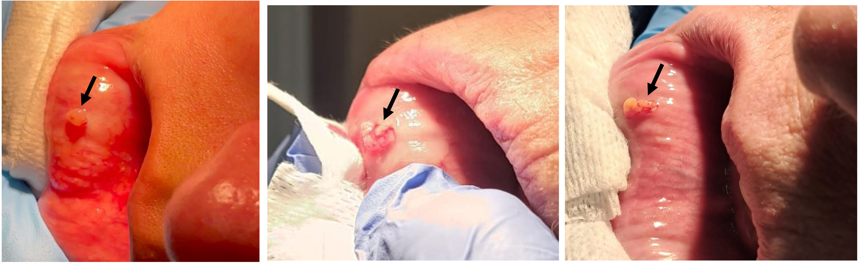
The doctor performing the procedure stands in front of the patient and identifies the salivary glands, which appear as small submucosal dots on the vestibular surface. The midline of the oral vestibule should be avoided, as it has a lower concentration of salivary glands. First, the local anaesthetic is injected into the oral mucosa, without going too deep, as the procedure is superficial. Secondly, a superficial vertical incision of 3−4mm is made with a scalpel. To facilitate the release of the gland, it is essential that the lip is taut; pressure can be applied to the contralateral side and to the sides of the incision (Fig. 2). The glands are separated from the underlying muscle layer with the tip of the scalpel, without enlarging the incision. Once they have been released and exposed on the lip (Fig.3), they are grasped with forceps, and their anchoring tissue is cut with the scalpel (Fig. 4). After the procedure, pressure is applied with sterile gauze for a few minutes until the bleeding stops (Fig. 5); if necessary, haemostatic gauze is applied. The sample is placed in a container with formalin and sent for processing to the Pathology Department (Fig. 6).
Sample processing


The sample is placed in a fixing solution: 10% neutral buffered formalin (pH 7.2–7.4). The container should ideally be made of plastic or clear glass and allow for the recommended 1:10 ratio of tissue to fixative (Fig. 6). It must be sent within a maximum of 24h, without the need for refrigeration during that time. It is then embedded in paraffin. Multiple tissue sections 4μm thick are made at 200μm intervals. It is then stained with haematoxylin and eosin, and the different cut levels are read, which enables the detection of foci in each section.29
ComplicationsWe have analysed the complications arising from MSGB performed using a minimal incision technique without sutures at our centre. To achieve this, we conducted a retrospective cross-sectional study using a telephone survey of patients who had undergone MSGB in the previous year. A total of 100 patients were included, most of whom were women (82%) with a mean age of 57±14 years. Eight patients (8%) were being treated with antiplatelet agents and four patients (4%) with anticoagulants, none of whom required discontinuation of treatment to undergo this technique. Of the total, only 24% had a minor complication and 9% had more than one. Minor complications included seven cases of self-limiting bleeding controlled with haemostatic gauze—only one of them in a patient on anticoagulants—six cases of self-limiting haematoma at 48h, and four cases of incision paraesthesia, of which only two persisted at the time of the telephone consultation. Ninety-eight per cent of the samples were valid for histological results.30
'Inconclusive' resultsEven when there is strong clinical suspicion, the results of MSGB can sometimes be inconclusive. In these cases, we may perform imaging tests such as ultrasound of the major and minor salivary glands and lacrimal glands, in addition to close clinical monitoring. Greyscale and Doppler ultrasound provide information on the involvement of the parotid and submandibular glands. Ultra-high-frequency ultrasound (over 20–30MHz, reaching 70MHz) obtains images with a resolution of up to 30μm of structures located within the first millimetre from the surface. This is a very attractive, non-invasive tool for assessing involvement of the labial salivary glands as it can identify early changes and guide and optimise a new biopsy.31 If the sample is not representative, we can repeat the biopsy within two to three weeks. If the inflammation is mild or not evident in the case of an incipient disease, we can repeat it later, at least after 12 months. Conversely, we may encounter cases where the gland is atrophic, for example in elderly individuals or in an advanced stage of the disease, with no inflammatory infiltrate.32
ConclusionsMSGB is useful beyond the classification and diagnosis of SS. It is decisive in diagnosing seronegative cases and is very valuable in patient’s follow-up, for example when there is suspicion of lymphoproliferative disease development or to monitor treatment response. It provides useful information on phenotypic stratification and prognosis, in terms of both severity and progression. Minimal incision techniques make MSGB an accessible tool for obtaining essential information and repeating the procedure during follow-up. Standardisation of its interpretation is necessary in certain clinical scenarios.
FundingThis article received no specific grant from any funding agency in the public, commercial, or not-for-profit sectors.
The authors have no conflict of interests to declare.