


Dengue is an infectious disease caused by the dengue virus (DV), which can progress to dengue hemorrhagic fever and dengue shock syndrome. DV causes the production of auto-antibodies against human cells. A variety of factors have been associated with macrophage activation syndrome, including infections, drugs and autoimmune pathologies (systemic lupus erythematosus, systemic onset juvenile idiopathic arthritis). We present three cases of patients that clinically developed an autoimmune response related to a DV infection. Our country currently has endemic cases of dengue, with hyperimmune responses. Therefore, the physician should consider this possibility in the presence of unusual conditions.
El dengue es una enfermedad infecciosa producida por el virus del dengue (VD), que puede evolucionar hacia fiebre hemorrágica dengue y síndrome de choque por dengue. El VD es causante de la producción de autoanticuerpos contra células humanas. Una amplia variedad de factores han sido asociados al síndrome de activación de macrófagos, incluyendo infecciones, fármacos y enfermedades autoinmunes de base (lupus eritematoso sistémico, artritis idiomática juvenil de inicio sistémico). Presentamos 3 casos de pacientes que desarrollaron clínicamente una respuesta autoinmune relacionada con una infección por VD. Nuestro país actualmente presenta casos de dengue en forma endémica, con respuestas hiperinmunes. Por lo tanto, el médico tratante debe pensar en esta posibilidad ante la presencia de afecciones no comunes.
Dengue is an infectious disease caused by the DV, which can progress to dengue hemorrhagic fever and dengue shock syndrome (DSS).1,2 Viral infection can also cause abnormal immune responses. Autoimmunity is characterized by autoantibody production and activation of autoreactive lymphocytes, which have been demonstrated to be associated to a number of viral pathogens. Previous studies indicate that the onset of the autoimmune response in dengue is a part of the pathogenesis of the disease; this can affect different organs and systems.3
We report 3 pediatric cases with autoimmune responses related to DV infection.
Case 1An 8-year-old male, with fever for 8 days, abdominal pain of 4 days of evolution in the epigastrium and right upper quadrant, which is accompanied by bloating, joint pain and general malaise. Physical examination: tachycardia, tachypnea, bilateral pleural effusion, globular abdomen, with collateral circulation, liver 8cm below the right costal ridge and spleen 12cm the left of the rib margin. The fever persisted after 14 days, with retroperitoneal lymphadenopathy. Lymph node biopsy was negative for malignancy. Serology for hepatitis, and Widal and Hudlesson reactions monotest, IgM isotype antibodies against toxoplasmosis, rK39 and IgM isotype antibodies vs CMV were all negative, IgG and IgM antibody isotypes for DV were positive. The cultures were negative. He also had proteinuria with hypoalbuminemia, bicytopenia (2500/ml leukocytes, platelets 80000/mm3) and hypocomplementemia. Lupus anticoagulant was negative, IgM isotype anticardiolipin antibodies were positive, C3 and C4 were normal, antinuclear antibodies (ANA) and anti-DNA were negative. Subsequently he presented improvement without treatment, and was discharged in good condition.
Case 2Male patient, 3 years old, with fever of 7 days, skin lesions for 7 days, predominantly on the trunk and upper limbs, erythematous macules, and petechiae of universal distribution. He also had abdominal pain of 2 days duration. Physical examination: tachycardia, tachypnea, petechiae. Bilateral breath sounds were rough, decreased in the right base. Abdomen globose, liver 3cm below the right costal margin and spleen 2cm below the left costal margin. Chest X ray showed condensation on the right base. By means of echocardiography, a minimal pericardial effusion was detected. Abdominal ultrasound found hepatosplenomegaly and free fluid in the abdomen. The dengue NS1 antigen was positive. The patient had persistent fever for 3 weeks with hepatomegaly 9cm below the right costal ridge and spleen 3cm below the left rib margin, with jaundice. Serology for hepatitis and the IgM isotype antibodies against CMV, Epstein–Barr virus, toxoplasmosis and HIV were all negative. Febrile antigens, ANA, anti-DNA and rheumatoid factor (RF) were also negative. The bone marrow aspirate was normal. Macrophage activation syndrome was diagnosed due to fever, leucopenia with neutropenia (3000/ml leukocytes, neutrophils 900/ml), anemia (hemoglobin 8.2g/dl), hepatosplenomegaly, triglycerides 470mg/dl and 1150 hyperferritinemia mg/dl. Boluses of methylprednisolone were initiated, which led to an improvement of the clinical status and the jaundice disappeared, as did the enlarged organs and the fever. He was discharged later.
Case 3Male infant, 3 months of age with fever for 4 days of evolution, in addition to vomiting and diarrhea 2 days earlier. Physical examination: globose abdomen, liver 3cm below the right rib margin. An abdominal ultrasound showed hepatosplenomegaly, thickened gallbladder wall and free fluid in the abdomen. The dengue NS1 antigen was positive. He had anemia (hemoglobin 8.0g/dl) which increase progressively with increasing hepatomegaly, up to 6cm below the right costal margin. Serology for hepatitis and IgM isotype antibodies against CMV, Epstein–Barr virus, toxoplasmosis and HIV were negative. Febrile antigens, ANA, anti-DNA and RF were negative. The marrow aspirate was normal. A diagnosis of macrophage activation syndrome was made due to fever, hepatosplenomegaly, leucopenia, neutropenia (3500/ml leukocytes, neutrophils 1100/ml), anemia (hemoglobin 8.0g/dl), hypertriglyceridemia 383mg/dl and hyperferritinemia 3828mg/dl. Boluses of methylprednisolone were started, with improvement of clinical status, decreased visceromegalies and hematologic improvement. He was subsequently discharged.
DiscussionIt is currently considered that there are several mechanisms that could explain the association of autoimmunity and viral infection, including molecular mimicry, cell activation and viral persistence. Previous studies showed that the DV infection leads to the production of autoantibodies against human cells. DV is an important infectious disease in tropical and subtropical regions of the world.1–4
The pathogenic mechanisms of dengue hemorrhagic fever (DHF) and DSS caused by DV infection remain unresolved. Patients with DV/DSS are characterized by various manifestations including severe thrombocytopenia, vascular leakage and hepatomegaly. Besides the effects of the viral load and the variation of the virus, abnormal host immune responses after DV infection may also account for the progression of DHF/DSS. Actually, autoimmunity is implicated in the pathogenesis of many viral infections such as HIV, hepatitis C, CMV, herpes simplex virus, Epstein–Barr and DV. Antibodies directed against the DV 1 nonstructural protein (NS1) show cross-reactivity with human platelets and endothelial cells, which leads to damage to the endothelial cells and platelets, as well as inflammation. These results, as well as the hypothesis that the anti-DV NS1 is involved in the pathogenesis of dengue and DHF/DSS can lead to important information on the development of a vaccine against dengue.5–8
A wide variety of factors have been associated with macrophage activation syndrome. These include infections (Epstein–Barr, varicella, Coxsackie, parvovirus B19, hepatitis A, Salmonella, Pneumocystis jiroveci, enterococci, leishmaniasis, etc.), drugs (aspirin, NSAIDs, methotrexate, etanercept, anakinra, gold salts, sulfasalazine and morniflumate) and autoimmune diseases (systemic lupus erythematosus, juvenile idiopathic arthritis of systemic-onset).9–13
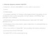
Hemophagocytic lymphohistiocytosis (HLH), which has many genetic causes, is characterized by multisystem inflammation. HLH is a reactive process resulting from prolonged and excessive activation of antigen presenting cells (macrophages, histiocytes) and CD8+T cells. Hemophagocytosis, which is mediated through CD163, is a characteristic of activated macrophages and histiocytes, giving the disease its name. The predominant clinical features of HLH are fever (often persistent), cytopenias, hepatitis and splenomegaly. The diagnostic protocol classification of HLH (Table 1) is used. Because of the involvement by the disease and life-threatening manifestations, treatment should be started as soon as possible with anti-inflammatory therapy, consisting of glucocorticoids, cyclosporine, etoposide or anti-thymocyte globulin (ATG). Secondary MAS is associated with autoimmune diseases or viral infections, with a significant mortality rate.11,14
Guidelines for the Diagnosis of Hemophagocytic Lymphohistiocytosis: HLH 2004 Protocol.
| 1. Molecular diagnosis consistent with HLH |
| 2. Diagnostic criteria for HLH fulfilled (5 or more of the 8 criteria below) |
| (a) Fever |
| (b) Splenomegaly |
| (c) Cytopenias (affecting 2 of 3 lineages in the peripheral blood): hemoglobin <9.0g/l (in infants less than 4 weeks: hemoglobin <10.0g/l), platelets <100000/mm3 three neutrophil count <1000/mm3 |
| (d) Hypertriglyceridemia and/or hypofibrinogenemia: triglycerides ≥265mg/dl, fibrinogen ≤1.5g/l |
| (e) Hemophagocytosis in bone marrow, spleen, lymph nodes, or cerebrospinal fluid: no evidence of malignancy |
| (f) Low or no activity of “natural killer” cells (according to the reference laboratory) |
| (g) elevated serum ferritin (≥500mg/l) |
| (h) soluble CD25 over the normal range for age |
HLH diagnosis can be performed with one or both criteria described in the table.
Our patients clinically presented an autoimmune response developed due to DV infection, which in the first case was self-limiting. The other 2 cases presented as secondary MAS and showed a good response to glucocorticoids. It is noteworthy that the last 2 cases showed no bone marrow hemophagocytosis, but met other criteria for the diagnosis in question.
Our country currently has cases of endemic dengue with hyperimmune responses. Therefore, the treating physician should consider this possibility in the presence of uncommon conditions.
Ethical ResponsibilitiesProtection of human and animal subjectsThe authors declare that no experiments have been performed on humans or animals.
Confidentiality of dataThe authors declare that they have followed the protocols of their workplace on the publication of data from patients and all patients included in the study have received sufficient information and gave written informed consent to participate in the study.
Right to privacy and informed consentThe authors have obtained informed consent from patients and/or subjects referred to in the article. This document is in the possession of the corresponding author.
Conflicts of InterestThe authors have no conflicts of interest.
Please cite this article as: Morel Z, Ramírez A. Respuesta autoinmune en niños con dengue. Reporte de casos. Reumatol Clin. 2014;10:257–259.


