

The appearance of inflammatory skin lesions (ISL), induced by biological drugs, mainly cutaneous psoriasis, has been extensively described.1–3 The hypothesis is that in patients with genetic susceptibility present an activation of alternative inflammatory pathways such as interferon-α 1.2, but this has not been demonstrated and no genetic studies in these patients exist.
We conducted a prospective observational study analyzing genetic data from patients of the Rheumatology Department of the Hospital del Mar (Barcelona) who developed de novo ISL due to biological therapy between January 2008 and December 2012.
The demographic and clinical variables evaluated were: age, sex, diagnosis of ISL, rheumatic disease and duration (in years), biologic drug employed and time from onset to the start of the drug (weeks). Genetic variables were the presence of HLA-B27, HLA-DR1, HLA-DR4 and HLA-DR7 alleles (detected by standard PCR), HLA-CW6 (Sanger sequencing using the indirect marker rs4406273) and the deletion of two genes late cornified envelope (LCE), LCE3C_LCE3B-del (with a multiplex PCR experiment4). Fifteen patients who developed ISL (prevalence 2.5%) were included. They had a mean age of 43.9±12.4 years and 73.3% were female. The diagnoses of ISL were: seven skin psoriasis (five palmoplantar pustulosis, one with psoriasis vulgaris and one with guttata), three alopecia areata, two cutaneous lupus, one eczema, one suppurative hidradenitis and one with erythema multiforme. Rheumatological diagnoses were: three rheumatoid arthritis, ankylosing spondylitis 6, four non-radiological axial spondyloarthritis and two psoriatic arthritis, mean disease duration of 13.1±7.4 years. Biologic drugs were eight cases using adalimumab, etanercept in four, infliximab in two and abatacept in one, with a mean time of onset of 57.1±62.1 weeks.
Genetically, seven patients had HLA-B27, four were HLA-CW6 positive (26%), only one with psoriasis, one positive HLA-DR1, another HLA-DR4 and six HLA-DR7 positive patients (40%), three with psoriasis. In four patients (26.7%) two LCE3C-LCE3B deleted alleles were detected and 11 (73.3%) had one deleted allele (Table 1). LCE3C_LCE3B allele frequency was 63.6%.
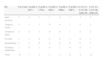
Genetic Characteristics of Patients with Cutaneous Lesions Induced.
| ISL | N.p.Total | N.p.HLA-B27+ | N.p.HLA-CW6+ | N.p.HLA-DR1+ | N.p.HLA-DR4+ | N.p.HLA-DR7+ | LCE 3C-LCE 3B-delD+/D+ | LCE 3C-LCE 3B-delD+/D− |
|---|---|---|---|---|---|---|---|---|
| Skin psoriasis | 7 | 4 | 1 | 1 | 0 | 3 | 3 | 4 |
| Alopecia areata | 3 | 1 | 1 | 0 | 0 | 1 | 1 | 2 |
| Cutaneous lupus | 2 | 0 | 1 | 0 | 0 | 1 | 0 | 2 |
| Eczema | 1 | 1 | 0 | 0 | 0 | 1 | 0 | 1 |
| Hydradenitis | 1 | 1 | 0 | 0 | 0 | 0 | 0 | 1 |
| Erythema multiforme | 1 | 0 | 1 | 0 | 1 | 0 | 0 | 1 |
| Total | 15 | 7 | 4 | 1 | 1 | 6 | 4 | 11 |
LCE 3B-3C-LCE del, deletion of the late cornified envelope; ISL, induced skin lesions; N.p., number of patients; +, positive; D+, deleted; D−, non-deleted.
The low proportion of patients with the presence of HLA-CW6 and HLA-DR7 alleles (especially in cases of psoriasis) stands out, lower than that found in populations of cutaneous psoriasis,5–7 which could be due to the predominant type of psoriasis, palmoplantar pustulosis, since the average age of patients was older than 40 years (HLA-CW6 and HLA-DR7 alone have been associated with type I psoriasis, with an onset before 40 years of age, and with the vulgaris and guttate subtypes5).
In our series, the presence of the deletion of the two LCE genes (allele frequency of 63.6%) was consistent with the frequently seen in population samples with inflammatory diseases (62%–70%) but higher than that reported in control populations (55%–60%).4,8–10 But what stands out is that all patients with ISL had at least one copy of the LCE3C_LCE3B deleted alleles (absence of non-deleted homozygous individuals). In the general population, the frequency of non-deleted homozygotes is around 18%4,9,10 and disruption of the skin barrier occurs in relation to the number of copies of LCE3C_LCE3B, being undetectable in carriers of the homozygous deletion4 and reduced in heterozygotes.
This observational study of a series of cases of ISL induced by biological drugs and is the first to include genetic data, although further studies are needed with larger numbers of patients and a control group to better study this process and help establish if there a pattern of genetic susceptibility as in the case of LCE3C_LCE3B deletions.
FinancingThe authors declare that there has been no financial support or commercial interest in carrying out this work.
Conflict of InterestThe authors have no conflict of interest to state.
Please cite this article as: Almirall M, Docampo E, Estivill X, Maymó J. Características genéticas de pacientes reumatológicos que desarrollan lesiones cutáneas inflamatorias inducidas por fármacos biológicos. Reumatol Clin. 2015;11:126–127.


