

We present the case of a 42-year-old male patient who was admitted for the study of axial and pelvic girdle pain, being diagnosed with hypophosphatemic osteomalacia secondary to a mesenchymal tumour after ruling out other causes of osteomalacia.
Se presenta el caso de un paciente varón de 42 años que ingresa para estudio de dolor axial y en cintura pélvica, siendo diagnosticado de una osteomalacia hipofosfatémica secundaria a tumour mesenquimal tras descartar otras causas de osteomalacia.
Oncogenic osteomalacia is a rare paraneoplastic syndrome. It is due to the overproduction of fibroblastic growth factor 23 (FGF-23) by small tumours, usually of mesenchymal lineage.1 FGF-23 produces hyperphosphaturia by inhibiting phosphate reabsorption in the renal proximal tubule,2 and as a consequence, hypophosphatemia and osteomalacia. To date, fewer than 1000 cases have been published in the literature.
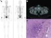
Clinical observationThe case concerns a 42-year-old male patient with no pathological history of interest. He was admitted to the Rheumatology ward for the study of dorsolumbar pain and bilateral pelvic girdle pain of more than one year of evolution that had been worsening in recent months, added to rib pain with progressive limitation in his usual activities. Laboratory tests on admission revealed a high alkaline phosphatase value (228 U/l [normal 38−126 U/l]), decreased phosphate (1.6 mg/dl [normal 2.5–4.5 mg/dl]) and a low value of 25-hydroxy-vitamin D (14.1 ng/ml [normal 20−100 ng/ml]). The remainder of the laboratory parameters were normal, including acute phase reactants and serum electrophoresis. In view of the polytopic bone pain, a bone scintigraphy with HDP-Tc99m of the whole body was requested (Fig. 1A), which showed the presence of multiple fracture foci in the costal arches, bony pelvis and left proximal femur. Magnetic resonance imaging of the spine and pelvis was also performed, identifying traces of fracture in both sacral wings, left iliopubic branch and at the pertrochanteric level of the left femur, as well as a slight wedge of T5, at the same time as identifying a lesion of kidney-like morphology of 32 × 22 mm in the right adductor region, with an increased signal in T2 and a hypointense centre. With these findings, hypophosphatemic osteomalacia was suspected and treatment with oral phosphate and cholecalciferol supplements was initiated. The study was completed with the measurement of FGF-23, which was elevated (348.2 IU/ml [normal < 180 IU/ml]), as well as an octreotide SPECT-CT scan that showed the presence of a mass in the deep right femoroinguinal region with moderate overexpression of somatostatin receptors (Fig. 1B). Given the presumptive diagnosis of oncogenic osteomalacia, the inguinal mass was surgically removed for characterisation. The pathological study confirmed the presence of a phosphaturic mesenchymal tumour (Fig. 1C). After surgery, serum phosphate and FGF-23 levels normalised, and within a few weeks the patient was asymptomatic.

A) Bone scan with multiple fractures: rib, bony pelvis and left proximal femur. B) SPECT-CT scan with octreotide-Tc99m showing right inguinal mesenchymal tumour with somatostatin receptor type 2. C) Histological section of the mesenchymal tumour (haematoxylin-eosin stain) with multinucleated osteoclast-like giant cells and haemosiderin deposits (haemosiderophages).
Hypophosphatemic osteomalacia secondary to a phosphaturic mesenchymal tumour is a rare pathology and is often underdiagnosed due to its insidious presentation and the difficulty in locating the causative tumour.1 In the case presented, we were able to observe clinical and biochemical resolution after total surgical resection. In the cases described, this entity is usually characterised by persistent hypophosphatemia, multiple bone fractures with associated pain, and proximal muscle weakness.3 The location of the tumour is crucial, since in most cases, when complete removal can be performed, the condition subsides entirely. The use of octreotide SPECT-CT has been shown to be a very useful localising technique for this type of tumour.4,5
ConclusionsThis case aims to illustrate the diagnostic challenges of this entity and to emphasize the importance of always including FGF-23-producing mesenchymal tumours in the differential diagnosis of hypophosphatemia.6
Ethical considerationsThe authors obtained the express agreement of the patient to publicly disseminate the clinical data and the results of additional tests provided.
The authors declare that there is no conflict of interest with respect to the information provided in this clinical case.


