Suggestions
Idioma
Guide for authors
Searcher
Journal Information
Visits
859
Vol. 21. Issue 9.
(November 2025)
Original article
Full text access

Impact of the IL18 −137 G/C (rs187238) polymorphism on susceptibility and clinical manifestations in women systemic lupus erythematosus

Impacto del polimorfismo IL18 -137 G/C (rs187238) en la susceptibilidad y las manifestaciones clínicas en mujeres con lupus eritematoso sistémico
Visits
859
Danton Magria,1, Clisten Fátima Staffena,1, Ticiana Della Justina Fariasb, Ilíada Rainha de Souzaa, Yara Costa Netto Muniza, Ivânio Alves Pereirac, Lia Kubelka de Carlos Backd, Luciano Santos Pinto Guimarãesa, Juliana Dal-Ri Lindenaua,
Corresponding author
juliana.lindenau@ufsc.br

Corresponding author.
a Genetic Polymorphisms Laboratory, Department of Cell Biology, Embryology and Genetics – Universidade Federal de Santa Catarina, Florianópolis, Brazil
b Department of Biological Sciences, University of North Carolina at Charlotte, Charlotte, NC, USA
c Rheumatology Division, University Hospital Polydoro Ernani de Sao Thiago, Federal University of Santa Catarina, Florianopolis, Brazil
d Biogenetika Individualized Medicine, Florianopolis, Brazil
This item has received
Article information
Abstract
Full Text
Bibliography
Download PDF
Statistics
Tables (3)
Table 1. Comparison between cases and controls to epidemiological variables.
Tables
Table 2. Estimation of the association between epidemiological variables and rs187238 with SLE susceptibility.
Tables
Table 3. Estimation of prevalence of clinical manifestations in *C carriers compared to the *GG genotype of the rs187238 polymorphism.
Tables
Additional material (1)
Abstract
Introduction and objectives

Systemic lupus erythematosus (SLE) is a chronic autoimmune disease characterized by the production of autoantibodies, inflammation processes, and tissue damage. There are several genetic factors associated with the disease, many of them single nucleotide polymorphisms (SNPs). Interleukin-18 is a pro-inflammatory cytokine encoded by the IL18 gene, and the SNP −137 G/C (rs187238) has been studied in several populations. This case control study analyzed whether rs187238 is associated with SLE susceptibility and its clinical manifestations in a Brazilian population.

Materials and methods

153 patients fulfilling the American College of Rheumatology classification criteria for SLE were recruited, as well as 147 controls. Genotyping was performed by sequence-specific polymerase chain reaction (SSP-PCR). To assess SLE susceptibility a logistic regression test was conducted. Clinical aspects were tested through Poisson regression and clustered by Principal Component Analysis.
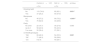
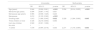
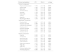
Results

An association between the rs187238*C_ carriers genotypes and SLE was found, these genotypes were associated with a 127% increased chance of developing the disease (OR=2.27, 95% CI=1.32–3.98, p=0.003). The *C_ genotypes were also associated with photosensitivity (PR=1.39, 95% CI=1.1–1.8, p=0.017), malar rash (PR=1.37, 95% CI=1.1–1.8, p=0.014) and Raynaud phenomenon (PR=1.37, 95% IC=1.1–1.8, p=0.015).

Discussion and conclusions

These findings suggest the potential of rs187238 as a genetic marker for SLE risk and clinical stratification in admixed Latin American populations.

Keywords:
Interleukin
Immunogenetics
Autoimmunity
Association study
Cytokine
Resumen
Introducción y objetivos

El lupus eritematoso sistémico (LES) es una enfermedad autoinmune crónica caracterizada por la producción de autoanticuerpos, procesos inflamatorios y daño tisular. Existen varios factores genéticos asociados con la enfermedad, muchos de ellos polimorfismos de un solo nucleótido (SNPs). La interleucina-18 es una citocina proinflamatoria codificada por el gen IL18, y el SNP -137 G/C (rs187238) ha sido estudiado en diversas poblaciones. Este estudio de casos y controles analizó si el rs187238 está asociado con la susceptibilidad al LES y sus manifestaciones clínicas en una población brasileña.

Materiales y métodos

Se reclutaron 153 pacientes que cumplían con los criterios de clasificación del Colegio Americano de Reumatología para LES, así como 147 controles. La genotipificación se realizó mediante reacción en cadena de la polimerasa con cebadores específicos de secuencia (SSP-PCR). Para evaluar la susceptibilidad al LES se realizó una regresión logística. Los aspectos clínicos se analizaron mediante regresión de Poisson y se agruparon mediante análisis de componentes principales.

Resultados

Se encontró una asociación entre los genotipos portadores del alelo C_ del rs187238 y el LES, estando dichos genotipos asociados con un aumento del 127% en la probabilidad de desarrollar la enfermedad (OR=2,27; IC95%: 1,32-3,98; p=0,003). Los genotipos *C_ también se asociaron con fotosensibilidad (PR=1,39; IC95%: 1,1-1,8; p=0,017), eritema malar (PR=1,37; IC95%: 1,1-1,8; p=0,014) y fenómeno de Raynaud (PR=1,37; IC95%: 1,1-1,8; p=0,015).

Discusión y conclusiones

Estos hallazgos sugieren el potencial del rs187238 como marcador genético de riesgo para LES y para la estratificación clínica en poblaciones latinoamericanas mestizas.

Palabras clave:
Interleucina
Inmunogenética
Autoinmunidad
Estudio de asociación
Citoquina
Full Text
Introduction

Systemic lupus erythematosus (SLE) is a chronic autoimmune disease, characterized by loss of self-tolerance, production of autoantibodies and multi-organ inflammation, including joint, skin, and renal. The clinical presentation of lupus is highly heterogeneous, occurring predominantly in women and the disease course is known by unpredictable flares and remissions.1 The interaction of genetic alterations with environmental factors and immunity (innate and adaptive) leads to increased immune complexes deposition, production of inflammatory cytokines and autoantibodies, along with organ damage. A chronic inflammatory state in SLE patients can result in morbidity and mortality.2

Single nucleotide polymorphisms (SNPs) have been reported to be associated with SLE susceptibility and may contribute to disease pathogenesis, including SNPs in the IL18 gene. IL18 is expressed by numerous immune cells and is known to be involved in multiple biological functions. The IL18 gene encodes the protein Interleukin-18 (IL-18), a pro-inflammatory cytokine that increases the activity of natural killer cells (NK) in the spleen and stimulates the production of gamma interferon (IFN-γ) by type I T-helper (Th) cells.3–5 Transcribed variants resulting from alternative splicing have also been reported. Its expression is increased in skin cells, esophagus and lymph nodes.

It is known that the SNP rs187238 located at the position −137 within promoter of the IL18 gene has three possible alleles, C>A and C>G, however, the *A allele is not usually observed in populations. The *G allele is associated with increased gene transcription and consequent greater production of the IL-18 protein. However, a few authors argue that, although this variant in the promoter region is associated with the production of pro-IL-18, it may not influence the release of biologically active IL-18.6

The heterozygous G/C genotype of this polymorphism has been associated with an increased susceptibility of developing arthritis in Swedish SLE patients.4 Besides, these authors suggested that the *C allele could reduce activity and IL-18 production. On the other hand, it has been reported that the *CC genotype was responsible for elevated IL-18 serum levels in Chinese SLE patients.7

Notwithstanding that several studies investigating this gene have been published, results from the association of IL18 SNPs with SLE susceptibility and its pathogenesis are still discrepant in different populations.8 Thus, how variations in this gene are associated with disease risk or clinical manifestations remains to be elucidated. Despite numerous studies in Asian and European populations, little is known about IL18 polymorphisms in admixed Latin American groups.4,7,8

Given IL-18's role in promoting Th1 immune responses and its elevated levels in SLE patients, genetic variants in its promoter may modulate disease risk. This study aimed to analyze the potential influence of IL18 gene polymorphism −137 G/C (rs187238) on genetic susceptibility to SLE and its association with distinct clinical features of the disease in a Brazilian population.

Materials and methodsPatients and healthy controls

This project was approved by the Ethics Committee on Human Research from Universidade Federal de Santa Catarina (trial number 423.535). A total of 153 women patients fulfilling the American College of Rheumatology (ACR) classification criteria for SLE were recruited, as well as 147 women controls without evidence of SLE or any other autoimmune disease nor any family cases. All patients answered a questionnaire regarding their symptoms and had hematological information collected from their medical records along with data from established inflammatory markers. Patients were consecutively recruited among the ones seeking care at the rheumatologic clinic from University Hospital Polydoro Ernani de São Thiago, Florianópolis, Brazil. Control participants were recruited from the same institution. Every participant signed an informed consent form (ICF) prior to blood drawn, authorizing the use of their samples as well as their clinical information in this study.

Clinical and laboratory data

Clinically relevant information such as smoking habit, menopause, and antibodies were categorized as present or absent. Parity was analyzed regarding the number of children (none; up to 2; 3 or more). Individuals’ ages as well as their menopause and menarche ages were treated as discrete variables. The results from laboratory exams were compared to standardized values, establishing normal and abnormal outcomes.

People with a smoking habit were divided between those who declared themselves to be smokers (who smoked regularly or had already smoked) and non-smokers (who had never smoked). Menopause status and parity were obtained through a questionnaire as well as menopause and menarche ages. The SLE clinical characteristics were obtained through medical records, based on the American College of Rheumatology criteria. Antibodies presence and laboratory exams were obtained through medical records since these are routine assays in this group of patients. All exams were conducted following gold standard techniques.

Genotyping

Genomic DNA was extracted from peripheral whole blood samples following the salting-out methodology.9,10 Genotyping was performed by sequence-specific polymerase chain reaction (SSP-PCR) and subsequently separated on a 2% agarose gel, following a protocol previously tested by other authors.11 An LPL gene fragment, flanking the polymorphic PvuII site in the intron between exon 6 and 7 of the gene was used as internal positive control.12 The polymerase, reagents and solutions concentrations used in this study are described in Supplementary Table S1. The primers sequences are described in Supplementary Table S2.

Statistical analysis

Allele and genotype frequencies were obtained through direct counting. Frequencies deviations were analyzed for the Hardy-Weinberg Equilibrium in both groups. Genotypes were grouped according to a frequency criterion, in which the low frequency categories were grouped (*C carriers versus *GG). Categorical variables were represented by absolute and relative frequencies. Significant differences between groups were accessed by comparison using a Chi-Square test with Yates’ correction when necessary. Continuous variables are presented as means±standard deviation (SD) or medians and interquartile range (IQR). Non-Gaussian variables were log-transformed for statistical analysis and reported after being back-transformed into their original units of measure. Student's t test was used for comparisons between groups.

The association of the SNP with SLE as well as epidemiological variables (smoking habit, menopause, parity, age, menopause and menarche ages) with SLE was determined through univariable logistic regression, estimating odds ratios (OR) and 95% confidence interval (CI). All variables with a p<0.1 in the univariable analysis were analyzed in a multivariable logistic regression. Age and smoking habit were used as covariates to perform the final model. The association between clinical data and SLE was examined by Poisson regression with robust variance, estimating prevalence ratios (PR) and 95% CI. Finally, a principal component analysis (PCA) was conducted to summarize patients’ clinical data into a smaller set of variables, therefore, three new variables were defined: “Clinical”, “Exams” and “Antibodies”. These three new variables were analyzed by Poisson regression with robust variance, estimating prevalence ratios (PR) and 95% CI to stablish the role of the polymorphism on SLE clinical progression. All analyses were performed with SPSS for Windows v. 25.0 (SPSS Inc., Chicago, IL, USA). Data were significant at p<0.05.

Results

The genotypic frequencies in both samples are in Hardy-Weinberg Equilibrium (data not shown). The allelic frequencies observed to each population are G=0.72 and C=0.28 in controls and G=0.54 and C=0.46 in SLE patients. Table 1 presents the univariable analysis of the association between epidemiological variables and SLE. A higher mean age was observed in the control group (49.1 years versus 37.9 in the SLE group) and this group showed more women in menopause (62.8% versus 26.8 in the SLE group). The SLE group had a higher percentage of smokers (37.9% versus 25.2% in the control group). Regarding the genotype distribution of rs187238, a higher frequency of the *CC genotype was observed in the SLE group compared to the control group. Additionally, the frequency of the *C allele carriers (*GC+*CC) was also increased among individuals with SLE (64.7%) compared to controls (53.7%), suggesting a possible association between the *C allele and susceptibility to SLE.

Table 1.

Comparison between cases and controls to epidemiological variables.

  Control (n=147)  SLE (n=153)  p-Value 
  n (%)  n (%)   
Smoking habit
No  110 (74.8)  82 (62.1)  0.031a
Yes  37 (25.2)  50 (37.9) 
Menopause
No  45 (37.2)  90 (73.2)  <0.001a
Yes  76 (62.8)  33 (26.8) 
Parity
No child  22 (16.7)  35 (27.6)  0.065
Up to 2  58 (43.9)  55 (43.3) 
3 or more  52 (39.4)  37 (29.1) 
rs187238 genotypes
*GG  68 (46.3)  54 (35.3)  0.001
*GC  71 (48.3)  59 (38.6) 
*CC  8 (5.4)  40 (26.1) 
  Mean (SD)  Mean (SD)  p-Value 
Age (years)  49.1 (14.9)  37.9 (12.4)  <0.001b 
Menarche age (years)  13.1 (1.7)  13.1 (1.8)  0.931 
  Median (IQR)  Median (IQR)  p-Value 
Menopause age (years)  49.0 [44.0; 52.0]  45.0 [42.0; 49.0]  0.052 

In bold are the variables with statistical significance at p<0.05.

a

Chi square test with Yates’ correction.

b

t test for independent samples. SD=standard deviation; IQR=interquartile range.

Table 2 shows the epidemiological and genetic variables analyzed through logistic regression considering the univariable and multivariable models. In the univariable analysis, age, menopause status, smoking habit, number of children, and polymorphism were selected based on the p-value<0.1 cutoff established, and they were inserted in the multivariable model. Considering the variable age, it was seen that the increase of one year provides around 7% protection against developing SLE. Regarding the smoking habit, individuals who smoke have around a 123% increased chance of developing SLE. Finally, when verifying the association with the −137 G/C polymorphism, it was observed that having the *C allele increases the individual's chance of having SLE by 127%. This result is maintained when adjusted for age and smoking habit in the multivariate analysis.

Table 2.

Estimation of the association between epidemiological variables and rs187238 with SLE susceptibility.

  UnivariableMultivariable
  OR  95% CI  p value  OR  95% CI  p value 
Age (years)  0.944  [0.926; 0.961]  <0.001  0.934  [0.914; 0.953]  <0.001 
Menarche age (years)  0.994  [0.867; 1.140]  0.931       
Menopause age (years)  0.965  [0.908; 1.025]  0.240       
Menopause  0.212  [0.122; 0.362]  <0.001       
Smoking habit  1.813  [1.089; 3.040]  0.022  2.228  [1.264; 3.990]  0.005 
Three children or more  0.435  [0.218; 0.850]  0.063       
Up to two children  0.580  [0.301; 1.099]         
No child  –  –       
*C allele  1.578  [0.994; 2.516]  0.053  2.271  [1.318; 3.984]  0.003 

Bold values represent statistical significance. The cut-off point for the inclusion of variables in the multivariable model was p=0.1; CI: confidence interval; OR: odds ratio; C: cytosine allele; SLE: systemic lupus erythematosus.

Clinical variables were analyzed through Poisson regression and the results were expressed as prevalence ratio (PR) (Table 3). Such a result is understood as the prevalence of a certain variable in SLE patients genotyped with the *C allele over the prevalence of this variable in patients having *GG genotype. Besides, the groups formed for PCA analysis are separated, through which significant results were obtained only for “Clinical” and “Exams”. Supplementary Table S3 shows the relation between each clinical variable and the analyzed polymorphism.

Table 3.

Estimation of prevalence of clinical manifestations in *C carriers compared to the *GG genotype of the rs187238 polymorphism.

Clinical manifestation  PR  95% CI  p-Value 
Clinical” component
Raynaud phenomenon  1.374  [1.1; 1.8]  0.015 
Discoid rash  1.225  [1.0; 1.5]  0.086 
Malar rash  1.368  [1.1; 1.8]  0.014 
Photosensitivity  1.388  [1.1; 1.8]  0.017 
Serositis  1.158  [0.9; 1.5]  0.245 
Oral ulcers  1.027  [0.8; 1.3]  0.825 
Arthritis  1.260  [0.9; 1.7]  0.160 
Nephritis  1.207  [1.0; 1.5]  0.108 
Neurological effects  1.078  [0.8; 1.4]  0.543 
Antibodies” component
Positive ANA  0.967  [0.7; 1.4]  0.864 
Positive anti-DNA  0.968  [0.7; 1.3]  0.820 
Positive anti-cardiolipin  1.194  [0.9; 1.6]  0.191 
Positive anti-LA  1.005  [0.7; 1.4]  0.976 
Positive anti-RNP  0.812  [0.6; 1.1]  0.143 
Positive anti-RO  1.034  [0.8; 1.3]  0.787 
Positive anti-Sm  0.973  [0.7; 1.4]  0.882 
Exams” component
Anemia  0.848  [0.7; 1.1]  0.202 
Leukopenia  0.816  [0.6; 1.1]  0.244 
Abnormal CRP  0.725  [0.6; 1.0]  0.020 
Abnormal ESR  0.984  [0.7; 1.3]  0.909 
Low hematocrit  0.675  [0.5; 0.9]  0.001 
High blood pressure  1.077  [0.8; 1.4]  0.562 
High LDL  1.198  [0.8; 1.7]  0.306 
High HDL  1.241  [0.8; 2.0]  0.377 
Elevated triglycerides  1.008  [0.7; 1.5]  0.972 
Thrombocytopenia  0.885  [0.6; 1.3]  0.507 

Bold values represent statistical significance at p<0.05. RP: prevalence rations; CI: confidence interval; SLE: systemic lupus erythematosus; ANA: antinuclear antibodies; DNA: deoxyribonucleic acid; CRP: C reactive protein; ESR: erythrocyte sedimentation rate; LDL: low-density lipoprotein; HDL: high-density lipoprotein.

Significant difference was observed in three variables when assessing clinical factors observed in SLE patients according to the polymorphism genotype: Raynaud's phenomenon, malar rash and photosensitivity, all more prevalent in individuals with the *C allele. Interestingly, results for C-reactive protein (CRP) reveal a greater number of patients with *GG genotype having elevated levels of this biomarker. This genotype was also more prevalent in patients with low hematocrit.

To better understand the relationship between the polymorphism and the clinical heterogeneity of SLE, a principal component analysis (PCA) was conducted. This multivariate technique allowed us to reduce the dimensionality of the dataset and cluster clinical and laboratory variables into three major components: “Clinical” (e.g., cutaneous and vascular manifestations), “Exams” (e.g., hematological and inflammatory markers), and “Antibodies” (e.g., ANA, anti-DNA, anti-Sm). The association between the *C allele and these components revealed that carriers were more likely to exhibit the clinical phenotype, while GG homozygotes showed more frequent alterations in laboratory exams, suggesting a differential impact of the polymorphism on disease expression

Considering the PCA results, regarding the group “Clinical”, individuals with the *C allele have a greater chance, about 20.7% (PR 1.207; CI 1.068–1.364; p=0.003), of having such phenotypes than people who are homozygous *GG. For the “Exams” group, patients with the *C allele are 17.1% (PR 0.829; CI 0.692–0.993; p=0.041) less likely to have altered results in these tests compared to patients with *GG genotype. Nonsignificant relation was observed in the “Antibodies” group (PR 0.967; CI 0.859–1.088); p=0.572). A fourth group was formed only including the significant “Clinical” variables for Poisson regression (Raynaud's phenomenon, malar rash, and photosensitivity) and it was observed that individuals with the *C allele are 27.2% (PR 1.272; CI 1.111–1.457; p=0.001) more likely to have such clinical outcomes than those homozygous *GG.

Discussion

In this case-control study of Brazilian women with SLE, we identified the IL18 −137*C allele as a risk factor not only for disease susceptibility but also for a distinct clinical profile. This is the first study that evaluated a Brazilian group of SLE patients and the polymorphic site −137 G/C of the IL18 gene to reveal association with susceptibility to the disease as well as certain clinical manifestations.

Regarding the association of SLE with the −137 G/C polymorphism, the logistic regression analysis revealed that this allele was more prevalent in patients than in controls, suggesting that having the *C allele increases a Brazilian individual's susceptibility of having SLE. A similar result was obtained by another group in a study with the Indian population.8 Besides, a *CG genotype on this site was related to an increased susceptibility of developing Hashimoto thyroiditis compared to a *GG genotype.13 The *C allele was also a susceptibility factor in type 1 diabetic patients with older age at onset (>15 years).14 Considering that these diseases are some of the most frequent autoimmune diseases, one could hypothesize that the *C allele at the −137 site may be a susceptibility factor to autoimmune diseases.

A meta-analysis reported a well-documented association for the SNP −137 G/C *GG genotype with SLE in the Asian population, but not in Europeans.15 This situation is an example of the patient's ancestry influence concerning genetic variations of the immune system. Other reviews did not even find an association of such SNP with SLE in a European population.16

Literature data concerning IL18 expression showed that the −137*C allele is involved in decreased IL-18 production on culture cells,4 suggesting that this allele destroys a histone 4 transcription factor-1 (H4TF-1) nuclear factor-binding site, resulting in a lower promoter activity.17 Recently, functional activity from IL18 promoter polymorphisms was analyzed by luciferase reporter assay and the SNP −137 G/C was able to modulate the transcriptional activity in the vector system. However, the authors discussed the incoherencies observed among expression studies concerning this gene promoter region and they attribute the variations to the fact that in vitro studies using isolated fragments of the regulating regions may not be able to correctly reflect the functional activity in vivo. Considering that cis- and trans-acting elements can act in IL-18 production, studies analyzing in vivo expression may help to clarify the role of these polymorphisms in modulating IL18 expression.18

It is well established that SLE patients have increased blood levels of IL-18, although the source remains unclear. It may be related to a genetic factor, which may play a vital role in SLE pathogenesis.19–21 This discrepancy between expression and availability can be partially explained by the bioavailability of IL-18: although individuals with a *GG genotype synthesize more, comparing to those with a *C carriers genotype, this molecule can be a pro-IL-18 and not necessarily the biologically active cytokine.6

Another two factors to be considered are (I) the several polymorphisms in this promoter region in high linkage disequilibrium that can generate different haplotypes and (II) the interaction of this gene with other cytokine genes, strongly influenced by the Brazilian ancestry of the studied sample. Considering that the linkage disequilibrium observed among the variants presented in this promoter region can vary depending on the analyzed population, it is important to consider the possibility to observe some association due to the ride effect.22 Furthermore, changes on linkage disequilibrium can affect the effect size of each variant in each population. These differences reinforce the need for population-specific studies, to understand the role of each marker in SLE susceptibility. Using genetic markers as disease biomarkers will only be possible with well-designed case controls studies in several different populations, such as the one presented here.

Assessing the clinical factors observed in SLE patients according to the polymorphism genotype, a significant difference was observed in three variables: Raynaud's phenomenon, malar rash and photosensitivity, all more prevalent in individuals with the *C allele. These findings are novel, as previous studies have not consistently linked the IL18 −137 G/C polymorphism to distinct clinical manifestations of SLE. Our results suggest that this variant may not only influence disease susceptibility but also modulate its phenotypic expression. It was observed that the *GG genotype on this locus in a northeastern Brazilian population affected by rheumatoid arthritis was related to lower disease activity.23 This study considered the −137IL18 site combined with several other polymorphisms in cytokine genes, demonstrating the importance of considering the role of a variant in its genomic context. Other authors also analyzed rheumatoid arthritis patients and verified that a lower percentage of extra-articular manifestations are present in *GG homozygotes when compared to other genotypes.24 Thus, the *C allele could be related to worst clinical parameters in rheumatoid arthritis patients, an important autoimmune disease, which converges with the results observed in this work for SLE pathogenesis. Our data suggest a potential role for the *C allele in SLE susceptibility, but further studies are necessary to disentangle the distinct contributions of the *GC and *CC genotypes observed in the literature, particularly in admixed populations such as ours. More studies also should be conducted to evaluate the possibility to use the variant rs187238 as a prognostic biomarker on autoimmune diseases.

The results for CRP are especially controversial. A greater number of SLE patients with *GG genotype had tests proving elevated levels of CRP comparing to those with the *C allele. Genome-wide association studies (GWAS) have shown that polymorphisms in the CRP gene causing its low expression are associated with SLE in various populations.25 CRP is mainly synthesized in the liver, during inflammation as a response to IL6 stimulation, thus being an acute-phase inflammation protein. The effects of low CRP levels in apoptotic cells and cellular debris clearance are currently under investigation; whether it plays a role in the pathogenesis of SLE remains to be elucidated.26 To date, no association has been reported between CRP and the IL18 gene.

The results for red blood cells ratio, expressed in the hematocrit test, revealed that a greater number of patients with *GG genotype have low hematocrit compared to those with the *C allele. A group has reported an association between low hematocrit and SLE patients, also observing that patients with such results had increased levels of CRP, suggesting a negative correlation between these two factors.27 Such results agree with those reported in this study, which shows that patients with high CRP also have low hematocrit, and this seems to be associated with the *GG genotype. It is estimated that 50% of SLE patients have kidney damage to some extent28 and this could reflect on hematocrit levels, since kidney impairment leads to low erythropoietin synthesis, which is a signal for decreasing erythrocyte production.

The principal component analysis revealed that individuals with the *C allele have a greater chance, about 20.7%, of having the clinical outcomes described in the “Clinical” group comparing to people who are homozygous *GG. Patients with the *C allele are also 17.1% less likely to have altered results in the tests included on the “Exams” group. Finally, the *C allele was linked to a 27.2% increased chance of having Raynaud's phenomenon, malar rash, and photosensitivity altogether. This analysis groups a great amount of information into smaller sets, making it easier to see the influence exerted by the SNP on the variables.

Despite having a low mortality rate, SLE symptoms usually greatly affect patients’ quality of life. Since the dominant characteristic of the disease is the production of autoantibodies and the exacerbated activation of inflammatory pathways, these patients suffer from chronic pain and fatigue, forcing them to continuously use anti-inflammatory drugs. Besides, any skin erythema drives them to adopt extra precautions regarding sun exposure.

Advances in molecular biology techniques have enabled scientists around the world to identify new loci associated with the disease, whether through GWAS or case-control studies. Such discoveries are motivated by knowing that complex conditions like SLE can be better understood when the number of patient data with different conditions, disease severity, and ancestry is increased. The results described here could potentially be used to integrate a genetic panel, improving SLE diagnosis. In addition, personalized medicine could take advantage of patients’ genotype for the IL18 −137 locus, given that initial studies showed better response on cloned cells for the *C allele regarding treatment with dexamethasone – a popular drug used to treat SLE.29

Differences in risk variants for autoimmune diseases exist between different populations and the prevalence of SLE varies substantially according to ethnic ancestry. Taking this into account, having conducted a research study addressing the SNP −137 G/C associated with SLE in women Brazilians is of great relevance, as it is a variant with scarce and often contradictory data. Acknowledging difficulties is the first step to perform and address results in an association study. The authors suggest further investigation to assess the role of the SNP in samples with varying ancestries, including different sets of Brazilian individuals, given the high diversification rates of this population, therefore improving reproducibility. It is important to highlight that the genetic and clinical heterogeneity of SLE across different populations may influence the association between rs187238 and disease susceptibility. Our study was conducted in Southern Brazil, a region characterized by a genetically admixed population resulting from European, African, and Amerindian ancestral contributions. Although ancestry inference was not performed in our sample, this genetic diversity may underlie differences in allele frequencies and disease expression. In this context, comparisons with other American populations, particularly those with Hispanic backgrounds, are highly relevant. Hispanic individuals in the United States and Latin America have been reported to present with earlier disease onset, more severe clinical manifestations, and poorer prognoses when compared to other ethnic groups.30 Given that genetic background modulates inflammatory pathways and cytokine expression, future studies should incorporate ancestry-informative markers or genome-wide data to better understand population-specific genetic risk factors and to assess whether the association of rs187238 with SLE varies across diverse genetic contexts.

The investigation of this type of polymorphism contributes to improve SLE treatment and diagnosis, additionally helping to elucidate disease aspects that remain unknown. These findings may contribute to the development of personalized strategies for clinical monitoring and treatment in genetically susceptible individuals. The rs187238 polymorphism, located in the promoter region of the IL18 gene, may influence transcriptional activity and IL-18 expression levels, which are central to inflammatory and immune pathways implicated in the pathogenesis of SLE. Although not yet established as a validated biomarker for clinical use, the determination of this genotype may offer insights into individual susceptibility and could be relevant for future strategies of risk stratification, particularly when considered in conjunction with other clinical and molecular factors. Given the multifactorial nature of SLE, it is important to acknowledge that the clinical impact of this polymorphism is likely to be modulated by complex interactions with epigenetic mechanisms – such as DNA methylation and histone modifications – as well as environmental exposures, including infections, UV radiation, and hormonal factors. These variables can influence cytokine expression patterns and immune dysregulation, contributing to the disease's heterogeneity in onset, progression, and therapeutic response.

Thus, while our data suggest a possible association between the *C allele of rs187238 and increased susceptibility to SLE, particularly in the studied population, this finding should be interpreted within the broader context of SLE pathophysiology. Future studies integrating genetic, epigenetic, and environmental data are warranted to elucidate the true clinical utility of this polymorphism and to determine whether it could serve as a useful adjunct in diagnosis, prognosis, or therapeutic decision-making in genetically diverse populations.

One of the limitations of this study is the absence of data from first-degree relatives of SLE patients. The inclusion of unaffected family members could provide valuable insights into the familial aggregation of the rs187238 polymorphism, helping to distinguish between inherited genetic susceptibility and gene-environment interactions that may contribute to disease onset. Investigating this variant in relatives would also allow for a more robust evaluation of its potential role in familial risk prediction and its segregation patterns within affected families. Future studies incorporating familial cohorts are warranted to explore these aspects and to clarify whether the observed increase in the *C allele frequency among patients is driven primarily by inherited predisposition or modulated by shared environmental exposures.

In conclusion, the IL18 rs187238 *C allele is associated with increased SLE susceptibility and with specific clinical features in Brazilian women. These results reinforce the need for population-specific genetic studies and suggest that rs187238 may serve as a potential biomarker for risk stratification and clinical monitoring in SLE.

Author contributions

DM, CFS were involved in data interpretation and manuscript writing. TDJF and YCNM contributed to study design and data collection. IRS, IAP and LKCB contributed to sample and data collection. LSPG contributed to study design, data analyses and data interpretation. JDL contributed to study design, data analyses, data interpretation and manuscript writing. All authors read and approved the final manuscript.

Conflicts of interest

The authors declare that there is no conflict of interest that could be perceived as prejudicial to the impartiality of the reported research.

Acknowledgements

We acknowledge the National Council for Scientific and Technological Development (CNPq/Brazil), the Coordination of Superior Level Staff Improvement (CAPES/Brazil, Finance code 001), the Graduate Program in Cell and Developmental Biology (PPGBCD/UFSC/Brazil), the Prorectorate for Research – PROPESQ (UFSC) and Universidade Federal de Santa Catarina (UFSC/Brazil) for the institutional support.

Appendix A
Supplementary data

The followings are the supplementary data to this article:

Icono mmc1.doc

References
[1]
B.L. Kotzin.
Systemic lupus erythematosus.
[2]
F. Favilli, C. Anzilotti, L. Martinelli, P. Quattroni, S. De Martino, F. Pratesi, et al.
IL-18 activity in systemic lupus erythematosus.
Ann N Y Acad Sci, 1173 (2009), pp. 301-309
[3]
H. Okamura, H. Tsutsi, T. Komatsu, M. Yutsudo, A. Hakura, T. Tanimoto, et al.
Cloning of a new cytokine that induces IFN-gamma production by T cells.
Nature, 378 (1995), pp. 703-706
[4]
V. Giedraitis, B. He, W.X. Huang, J. Hillert.
Cloning and mutation analysis of the human IL-18 promoter: a possible role of polymorphisms in expression regulation.
J Neuroimmunol, 112 (2001), pp. 146-152
[5]
S.A. Ihim, S.D. Abubakar, Z. Zian, T. Sasaki, M. Saffarioun, S. Maleknia, et al.
Interleukin-18 cytokine in immunity, inflammation, and autoimmunity: biological role in induction, regulation, and treatment.
[6]
K. Yasuda, K. Nakanishi, H. Tsutsui.
Interleukin-18 in health and disease.
Int J Mol Sci, 20 (2019), pp. 649
[7]
Q. Xu, K.T. Soe, S.S. Paramalingam, J. Thumboo, D.-R. Koh, K.-Y. Fong.
Interleukin-18 promoter gene polymorphisms in Chinese patients with systemic lupus erythematosus: association with CC genotype at position −607.
Ann Acad Med Singapore, 36 (2007), pp. 91-95
[8]
V. Umare, V. Pradhan, S. Nath, A. Rajadhyaksha, K. Ghosh, A.H. Nadkarni.
Impact of functional IL-18 polymorphisms on genetic predisposition and diverse clinical manifestations of the disease in Indian SLE patients.
Lupus, 28 (2019), pp. 545-554
[9]
D.K. Lahiri, J.I. Nurnberger.
A rapid no-enzymatic method for the preparation of HMW DNA from blood for RFLP studies.
Nucleic Acids Res, 19 (1991),
[10]
S.A. Miller, D.D. Dykes, H.F. Polesky.
A simple salting out procedure for extracting DNA from human nucleated cells.
Nucleic Acids Res, 16 (1998),
[11]
S.D. Celik, O. Ates.
Genetic analysis of interleukin 18 gene polymorphisms in alopecia areata.
J Clin Lab Anal, 32 (2018), pp. e22386
[12]
J.P. Johnson, P.M. Nishina, J.K. Naggert.
PCR assay for a polymorphic Pvull site in the LPL gene.
Nucleic Acids Res, 18 (1991), pp. 7469
[13]
D. Karakaya, Genc GÇ, S.K. Celik, T. Aktas, T. Bayraktaroglu, A. Dursun.
Association between IL-18 gene polymorphisms and Hashimoto thyroiditis.
Mol Biol Rep, 48 (2021), pp. 6703-6708
[14]
Z. Mojtahedi, S. Naeimi, S. Farjadian, G.R. Omrani, A. Ghaderi.
Association of IL-18 promoter polymorphisms with predisposition to Type 1 diabetes.
Diabetic Med, 23 (2006), pp. 235-239
[15]
G.G. Song, S.J. Choi, J.D. Ji, Y.H. Lee.
Association between interleukin-18 polymorphisms and systemic lupus erythematosus: a meta-analysis.
Mol Biol Rep, 40 (2013), pp. 2581-2587
[16]
B.R. Guo, J.J. Rong, Y.H. Wei, W.Y. Zhong, C.S. Li, M. Liu, et al.
Ethnicity-stratified analysis of the association between IL-18 polymorphisms and systemic lupus erythematosus in a European population: a meta-analysis.
Arch Dermatol Res, 307 (2015), pp. 747-755
[17]
S.B. Anyona, P. Kempaiah, E. Raballah, C. Ouma, T. Were, G.C. Davenport, et al.
Functional promoter haplotypes of interleukin-18 condition susceptibility to severe malarial anemia and childhood mortality.
Infect Immun, 79 (2011), pp. 4923-4932
[18]
A.U. AlRuwaisan, M.R. Al-Anazi, M.I. Shafeai, F.H. Rudiny, A.M. Motaen, S.M. Bin Dajem, et al.
Associations of single nucleotide polymorphisms in IL-18 gene with plasmodium falciparum-associated malaria.
J Inflam Res, 14 (2021), pp. 3587-3619
[19]
R. Mende, F.B. Vincent, R. Kandane-Rathnayake, R. Koelmeyer, E. Lin, J. Chang, et al.
Analysis of serum interleukin (IL)-1β and IL-18 in systemic lupus erythematosus.
[20]
Y.H. Lee, G.G. Song.
Circulating interleukin-18 level in systemic lupus erythematosus.
J Rheum Dis, 27 (2020), pp. 110-115
[21]
M. Xiang, Y. Feng, Y. Wang, J. Wang, Z. Zhang, J. Liang, et al.
Correlation between circulating interleukin-18 level and systemic lupus erythematosus: a meta-analysis.
[22]
J. Arimitsu, T. Hirano, S. Higa, M. Kawai, T. Naka, A. Ogata, et al.
IL-18 gene polymorphisms affect IL-18 production capability by monocytes.
Biochem Biophys Res Commun, 342 (2006), pp. 1413-1416
[23]
M.L. Monteiro, D.A. Barboza, E. Rushansky, M.H. Mariano, P. Sandrin-Garcia, P.R. De Souza, et al.
IL1β, IL18, NFKB1 and IFNG gene interactions are associated with severity of rheumatoid arthritis: a pilot study.
Autoimmunity, 53 (2020), pp. 95-101
[24]
A. Pawlik, M. Kurzawski, M. Drozdzik, V. Dziedziejko, K. Safranow, M. Hercynska.
Interleukin-18 gene (IL18) promoter polymorphisms in patients with rheumatoid arthritis.
Scand J Rheumatol, 38 (2009), pp. 159-165
[25]
S. Gaitonde, D. Samols, I. Kushner.
C-reactive protein and systemic lupus erythematosus.
Arthritis Care Res, 59 (2008), pp. 1814-1820
[26]
S.M. Nehring, A. Goyal, B.C. Patel.
C reactive protein.
StatPearls Publishing, (2021),
[27]
M. Yang, N. Ma, H. Fu, T. Wei, Q. Tang, B. Qin, et al.
Hematocrit level could reflect inflammatory response and disease activity in patients with systemic lupus erythematosus.
Clin Lab, 61 (2015), pp. 801-807
[28]
S. Almaani, A. Meara, B.H. Rovin.
Update on lupus nephritis.
Clin J Am Soc Nephrol, 12 (2017), pp. 825-835
[29]
J. Qiu, X. Qin, J. Wen, L. Wu, L. Kong, Y. Ou, et al.
Single-nucleotide polymorphisms influence the promoter activities of systemic lupus erythematosus-associated genes.
Biotechnol Lett, 42 (2020), pp. 1887-1896
[30]
G.S. Alarcón, G. McGwin Jr., M. Petri, J.D. Reveille, R. Ramsey-Goldman, R.P. Kimberly, et al.
Baseline characteristics of a multiethnic lupus cohort: PROFILE.

These authors contributed equally for this work.

Copyright © 2025. Sociedad Española de Reumatología (SER), Colegio Mexicano de Reumatología (CMR) and Elsevier España, S.L.U.
Download PDF
Idiomas
Reumatología Clínica (English Edition)
Article options
Tools
Supplemental materials