Journal Information
Vol. 10. Issue 1.
Pages 56-57 (January - February 2014)
Vol. 10. Issue 1.
Pages 56-57 (January - February 2014)
Images in Clinical Rheumatology
Full text access
Infectious Tenosynovitis of the Foot Extensors
Tenosinovitis infecciosa del aparato extensor del pie
Visits
16430
Carlos Guillén Astete
Corresponding author
, Ana Rodríguez García, Marta Prieto Morales, Antonio Zea Mendoza
Servicio de Reumatología, Hospital Universitario Ramón y Cajal, Madrid, Spain
This item has received
Article information
Full Text
Bibliography
Download PDF
Statistics
Figures (2)
Full Text

A 50-year-old male came to the emergency room with fever and pain in the left foot for the past 10h. He had no history of trauma.

On physical examination we found erythema, and warm, tender swelling of the distal forefoot with pain intensified by flexion of the third toe.

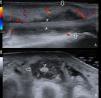
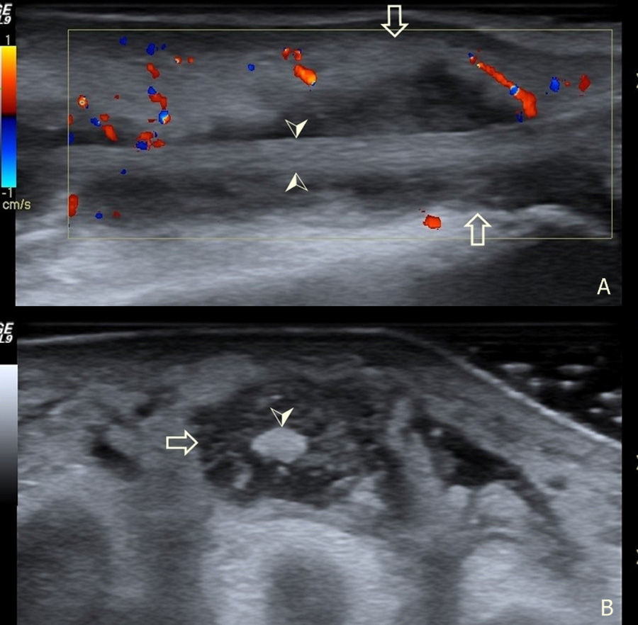
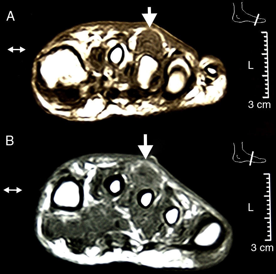
An ultrasound of the foot determined the presence of an hypoechoic effusion into the sheath of the extensor of the third toe at the middle and distal thirds with moderate color Doppler signal and no increase in skin thickness (Fig. 1). Since usually branches of the digitorum extensor have no sheath beyond the middle third of the forefoot1 we confirmed the diagnosis with an MRI (Fig. 2). We started antibiotic treatment with vancomycin from the first contact, and debridement was performed on the second day, draining 12cc of purulent-looking fluid from which we identified a strain of Staphylococcus aureus, which coincided with the results of blood cultures.

Fig. 1.

Ultrasound of the dorsal region of the left foot. (A) Longitudinal section of the third toe extensor. (B) Cross section of the same tendon. Hollow arrow: limits of the tendon sheath. Arrowhead: limits of the tendon within the sheath.

(0.11MB).
Fig. 2.

MRI of the foot in two cross sections: distal third in the upper image and middle third in the lower image. The effusion and hypertrophy of the sheath and the integrity of the cortical bone and lack of skin involvement can be observed.

(0.13MB).

Infectious tenosynovitis of the foot occurs most often at the plantar flexors and is related to cutaneous perforation by foreign bodies,2 a very rare presentation is on the foot extensor region. Mechanical tenosynovitis of the extensor tendons is usually associated with chronic exposure to repeated trauma3 that occurs in the ultramarathoner foot syndrome due to mechanical contact between the tendon and proximal osteophitic prominence wedges.4 Cellulitis of the dorsum of the foot is the first differential diagnosis. The diagnosis can be made through ultrasound5 visualizing the tendon sheath immersed in a hypoechoic fluid and confirmed through aspiration and microbiological study. MRI can also assess the existence of6,7 bone involvement. Antibiotic treatment should be started before the diagnostic suspicion, although the duration and route of administration is heterogeneous in the few similar case reports found.8–10 In our opinion, systemic compromise justifies intravenous antibiotic treatment.

Ethical Responsibilities

Protection of people and animals. The authors state that no experiments were performed on humans or animals.Data confidentiality. The authors declare that they have followed the protocols of their workplace regarding the publication of data from patients and all patients included in the study have received sufficient information and gave their written informed consent to participate in this study.Right to privacy and informed consent. The authors have obtained informed consent from patients and/or subjects referred to in the article. This document is in the possession of the author of correspondence.

Disclosures

The authors have no disclosures to make.

References
[1]
P. Hernandez, W. Hernandez, A. Hernandez.
Clinical aspects of bursae and tendon sheaths of the foot.
J Am Podiatr Med Assoc, 81 (1991), pp. 366-372
[2]
C.-H. Tung, Y.-H. Chen, H.H.C. Lan, T.-Y. Hsieh, D.-Y. Chen, J.-L. Lan.
Diagnosis of plant-thorn synovitis by high-resolution ultrasonography: a case report and literature review.
Clin Rheumatol, 26 (2007), pp. 849-851
[3]
W.D. Bruce, M.R. Christofersen, D.L. Phillips.
Stenosing tenosynovitis and impingement of the peroneal tendons associated with hypertrophy of the peroneal tubercle.
Foot Ankle Int, 20 (1999), pp. 464-467
[4]
H. Kobayashi, M. Sakurai, T. Kobayashi.
Extensor digitorum longus tenosynovitis caused by talar head impingement in an ultramarathon runner: a case report.
J Orthop Surg (Hong Kong), 15 (2007), pp. 245-247
[5]
O.S. Rasmussen.
Sonography of tendons.
Scand J Med Sci Sports, 10 (2000), pp. 360-364
[6]
C. Pineda, R. Espinosa, A. Pena.
Radiographic imaging in osteomyelitis: the role of plain radiography, computed tomography, ultrasonography, magnetic resonance imaging, and scintigraphy.
Semin Plast Surg, 23 (2009), pp. 80-89
[7]
C. Pineda, A. Vargas, A.V. Rodríguez.
Imaging of osteomyelitis: current concepts.
Infect Dis Clin North Am, 20 (2006), pp. 789-825
[8]
S. Faraj, D. Stanley-Clarke.
Acute extensor hallucis longus tenosynovitis caused by gonococcal infection.
N Z Med J, 116 (2003), pp. U421
[9]
R.A. Schaefer, R.J. Enzenauer, A. Pruitt, R.S. Corpe.
Acute gonococcal flexor tenosynovitis in an adolescent male with pharyngitis. A case report and literature review.
Clin Orthop Relat Res, 281 (1992), pp. 212-215
[10]
M. Nyska, A.D. Sperber, C.B. Howard, A. Nyska, S. Dekel.
Ankle extensor tendon synovitis due to a date palm thorn.
Foot Ankle, 10 (1989), pp. 180-183

Please, cite this article as: Guillén Astete C, et al. Tenosinovitis infecciosa del aparato extensor del pie. Reumatol Clin. 2014;10:56–57.

Copyright © 2012. Elsevier España, S.L.. All rights reserved
Idiomas
Reumatología Clínica (English Edition)
Article options
Tools
es en

¿Es usted profesional sanitario apto para prescribir o dispensar medicamentos?

Are you a health professional able to prescribe or dispense drugs?