


We report the case of a 59-year-old woman who presented with a mildly painful, bluish nodular lesion located in the connective tissue adjacent to the metacarpophalangeal joint of the first finger on her left hand. Soft tissue ultrasound (Fig. 1A and B) showed a turbulent vascular flow within the lesion, with a low-resistance arterial waveform and an afferent arterial branch that reached toward the lesion described above. Magnetic resonance imaging (MRI) (Fig. 1C) identified the lesion as being hyperintense (arrow) on a fat-saturated T2-weighted sequence. These findings suggested a vascular lesion and, when asked, the patient confirmed that she had had a prior traumatic injury. Thus, the possibility of a posttraumatic pseudoaneurysm or a hemangioma was considered. After surgical removal of the lesion, the pathological analysis of the resected specimen (Fig. 1D) showed numerous vascular spaces, surrounded by an endothelium in which nothing atypical was observed, lying on a stroma with hyalinized papillae. An immunohistochemical study with the anti-CD34 antibody confirmed the vascular differentiation of the lesion. The histopathological findings corresponded to an intravascular papillary endothelial hyperplasia, also known as Masson's hemangioma or Masson's tumor.1,2
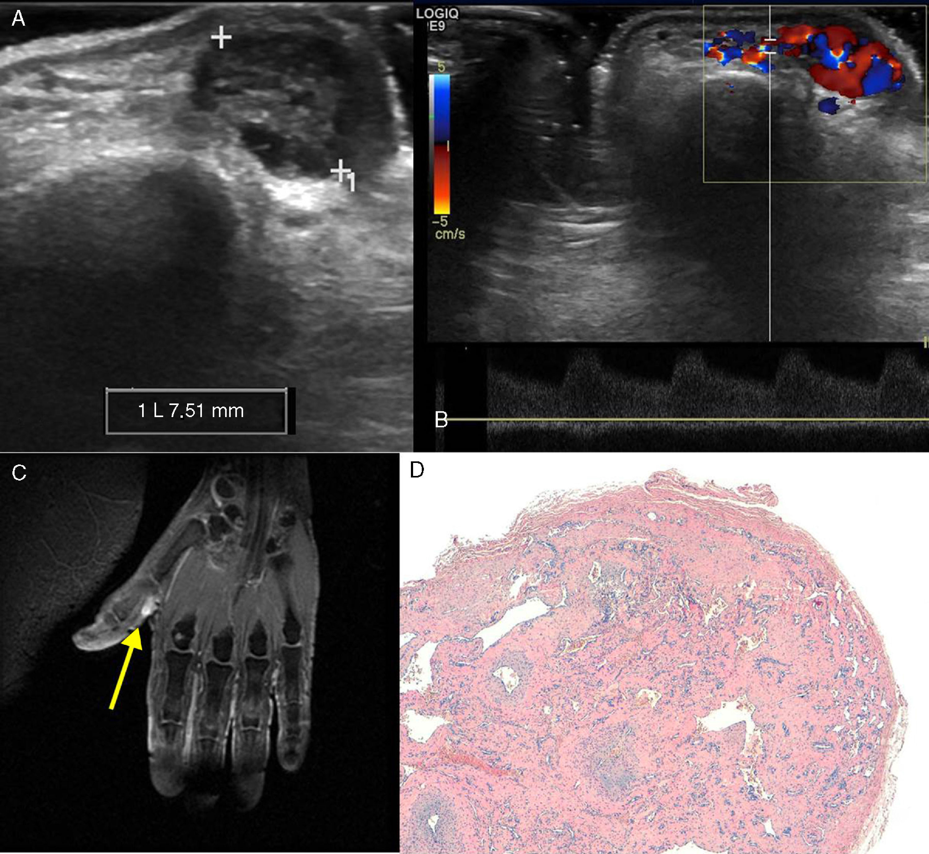
(A) Ultrasound image in B mode showing a 7.5-mm lesion in the soft tissue of the proximal phalanx of the first digit of the left hand. (B) Spectral Doppler ultrasound showing a low-resistance arterial waveform. (C) Fat-saturation T2-weighted magnetic resonance image showing the hyperintensity of the lesion (arrow). (D) The pathological study using hematoxylin–eosin staining revealed numerous vascular spaces surrounded by an endothelium with no atypical features, lying on a stroma with hyalinized papilla.
Intravascular papillary endothelial hyperplasia is a proliferative vascular lesion, generally formed in response to traumatic injury,3 that represents 2%–4% of the vascular soft tissue tumors.2,4 It occurs more frequently in women than in men,1,3 although its incidence does not differ according to age range or race.4 Three types have been distinguished on the basis of Hashimoto's classification: primary, which develops in dilated vascular spaces; mixed, which occurs in existing vascular anomalies, such as hemangiomas, arteriovenous malformations and pyogenic granulomas; and the third, a rare, extravascular type that develops in the interior of a hemotoma.2 The typical sites of this lesion are the subcutaneous vessels of the head, neck, fingers and trunk.1,2,4,5 The treatment of choice is complete surgical resection, because these masses do not usually resolve spontaneously.2
Ethical disclosuresProtection of human and animal subjectsThe authors declare that no experiments were performed on humans or animals for this study.
Confidentiality of dataThe authors declare that no patient data appear in this article.
Right to privacy and informed consentThe authors declare that no patient data appear in this article.
Conflicts of InterestThe authors declare they have no conflicts of interest.
Please cite this article as: Sánchez-Oro R, Sanchís-García JM, Saravia M, Bértolo-Domínguez M. Hiperplasia endotelial papilar intravascular: apariencia ecográfica y en resonancia magnética con correlación histopatológica. Reumatol Clin. 2016;12:167–168.


