


Examining cytokine profile complexities in chronic autoimmune disorders holds significant clinical importance. In order to address the similarities and differences related to SLE and RA, it was necessary to evaluate their cytokine chemokine profiles. Such analyses would give pointers towards differences, leading thereby to explore the potential of cytokines/chemokines as biomarkers. The study was therefore driven by the concept of understanding the major differences at this level with a hope of contribution towards diagnostics/theranostics. A multiplex study was carried out on systemic autoimmune disorders, such as SLE and RA, analysing forty analytes in comparison with healthy controls.
MethodologyAge and sex matched healthy donors and patients (n=38) were recruited and plasma cytokine profiling was done by Bio-plex multiplex immunoassay system.
ResultsA comparison with healthy volunteers revealed differential alteration in various chemokines in SLE and RA, respectively. Protein interaction analysis identified a core complex of chemokines (CXCL10, CCL5, CXCL12, CXCL9, CXCL1, and CXCL27) as central modulators, suggesting their potential as biomarkers. Drug prediction using the DSigDB database identified acetovanillone as a potential drug against this core complex. In comparing lupus patients with or without arthritis comorbidity, elevated levels of cytokines: IL-12, SCF, and TNF-a were prominently associated with arthritis in SLE. TNF-a emerged as a potential indicator specifically for arthritis.
ConclusionThis study enhances our understanding of the complex interplay of cytokine/chemokine in these systemic conditions and suggests their utility as targets and diagnostic paradigms for detection.
Examinar las complejidades del perfil de citocinas en los trastornos autoinmunes crónicos tiene una importancia clínica significativa. Con el fin de abordar las similitudes y diferencias relacionadas con el lupus eritematoso sistémico (LES) y la artritis reumatoide (AR), fue necesario evaluar sus perfiles de citocinas y quimiocinas. Tales análisis darían indicaciones sobre las diferencias, lo que conduciría a explorar el potencial de las citocinas/quimiocinas como biomarcadores. El estudio estuvo impulsado por el concepto de comprender las principales diferencias en este nivel con la esperanza de contribuir al diagnóstico/teranóstico. Se realizó un estudio multiplex en trastornos autoinmunes sistémicos, como el LES y la AR, analizando 40 analitos en comparación con los controles sanos.
MetodologíaSe reclutaron donantes sanos emparejados por edad y sexo y pacientes (N=38) y se realizó el perfilado de citocinas plasmáticas mediante el sistema de ensayo inmunológico multiplex Bio-Plex®.
ResultadosUna comparación con voluntarios sanos reveló una alteración diferencial en varias quimiocinas en el LES y en la AR, respectivamente. El análisis de interacción de proteínas identificó un complejo central de quimiocinas (CXCL10, CCL5, CXCL12, CXCL9, CXCL1 y CXCL27) como moduladores centrales, sugiriendo su potencial como biomarcadores. La predicción de medicamentos utilizando la base de datos DSigDB identificó acetovanillone como un posible medicamento contra este complejo central. Al comparar los pacientes con lupus con o sin comorbilidad de artritis, los niveles elevados de citocinas: IL-12, SCF y TNF-α estuvieron prominentemente asociados con la artritis en el LES. El TNF-α surgió como un indicador potencial específicamente para la artritis.
ConclusiónEste estudio mejora nuestra comprensión de la compleja interacción de citocinas/quimiocinas en estas condiciones sistémicas, y sugiere su utilidad como objetivos y paradigmas diagnósticos para la detección.
Systemic lupus erythematosus (SLE) and rheumatoid arthritis (RA) are autoimmune disorders of unknown etiology. The prevalence of SLE and RA is 5.14 and 0.75 per 100,000 person per year respectively.1 Systemic conditions in SLE can exhibit arthritis as comorbidity whereas RA is limited to specific regions of the body. Common susceptible factors are known to escalate both the pathologies, but may result in the development of different disease conditions. Immunobiology of both disorders interrelate which reveals the interplay of different immune cells and cytokines. Since the level of multiple cytokines increased among both the pathologies, therefore it is very difficult to differentiate the patients of both disorders on basis of their cytokine profiles. Thus, understanding the complex play of cytokine levels among these patients with different disease severity states can be of importance in classifying as well as in designing their therapeutic regimes.
SLE is a prototype of systemic autoimmune condition where the increased level of various cytokines can be observed in saliva, urine, and in various organs. The level of IL-2 was diminished in the lupus patients and anti-IL-2 antibodies were also observed to be associated with disease activity.2 IL-38 level was positively correlated with diseases activity and negatively correlated with C3 and C4 protein level.3 The level of IL-12 correlated positively with SLEDAI and could also divert the T cell development towards the Th1 lineage. IL-23 diverts the T cell development towards Th17 cells which leads to the secretion of IL-17, found to be coupled with lupus nephritis. A number of chemokines were also observed to have potent role in modulating the severity in SLE. CCL2/MCP-1, CCL11/eotaxin, CXCL1, CXCL8/IL-8, CXCL10/IP-10, CXCL12/SDF-1 levels in lupus patients were found to be coupled with kidney manifestation and also correlated with CNS manifestations.4 Heterogeneity among cytokine and chemokine levels in lupus patients can make it difficult to predict them as biomarker(s) and hence the need for multiplexing and further clustering of cytokines based on different disease stages.
Rheumatoid arthritis (RA) is a systemic disease, causing autoimmune response leading to joint damage and lifelong disability in severe conditions. RA can also exhibit in two states: active or inactive which can be importantly modulated by the interaction of cytokines. The two arms of immune system are facilitated by IFNs that modulate severity of RA. TNFs shows the dual role in RA pathology as it can protect against arthritic condition when secreted by the T cell or it can flare the conditions when secreted by the B cells.5 TNFs and IL-17 up regulations can further amplify the expression of various effector molecules such as CXCL1, CXCL2, CXCL3, IL-6, IL-8, and MMP-3.6 IL-6 also triggers the secretion of VEGF which is an important cytokine in RA inducing angiogenesis and immune cell migration into joints triggering inflammatory cascade. IL-17 can also trigger the secretion of RANKL, which is crucial for osteoclast differentiation. Inflamed joints have elevated level of RANKL which in turn increases the osteoclast differentiation and development of bone erosion in RA.7 Understanding of the complex cytokine network in RA will provide the insight into pathological mechanisms as well as highlight novel biomarkers of diagnostic importance.
Cytokine inhibitors such as those against TNFs and IL-6 have already proved to be of clinical importance for both the pathologies. GM-CSF inhibitors were also employed for better outcomes among RA patients. Recently, the treatment strategies are modifying by shifting towards the disease-modifying anti-rheumatic drugs (DMARDS) which have potential to inhibit the intracellular signalling induced by the extracellular cytokines. However, studies showing the complex interplay of the various cytokines creates the urgent need of a therapy with multiple targets for the cytokines. Advancement in molecular biology techniques such as multiplexing enables us in understanding of functional interaction among cytokines at different disease states. Several recent studies have also employed such strategies to understand the functional and correlational interaction among cytokines from autoimmune patients. In this study, we analysed 40 analytes including cytokines and chemokines among healthy control (HC), lupus patients (SLE) and arthritis patients (RA). Alterations in the cytokine profiles were compared between HC group vs SLE or RA. Arthritis comorbidities among SLE patients were also compared with RA patients. The functional and correlation interactions among cytokines in SLE and RA patients might provide the important diagnostic evidence as well as abnormal interactions within cytokine profiles.
Material and methodologyStudy design and sample collectionThe study was designed based on the case–control and cross disorder study where cytokine levels among various groups: SLE and RA patients with healthy controls; SLE and RA patients were compared. Arthritis comorbidities among lupus patients were also compared with cytokine profile of RA patients. Further, the observed differences among the cytokine profiles were utilised for the prediction of potential biomarkers as well as novel drug(s).
Age and sex matched healthy donors and patients (n=38) were recruited at post graduate institute of medical education and research (PGIMER), Chandigarh after obtaining an approval form ethics committee (IEC# IEC-05/2016-419 and IEC-01/2022-2277). The study was carried out in accordance with the ethics principle and all the subjects were made aware of the study and an informed consent was taken before sample collection. A set of inclusion and exclusion criteria were used for the selection of patients: women of age over 18 years and diagnosed with SLE or RA were included and pregnant women or those with any other disease were excluded from the study. All the patients were diagnosed according to the ACR guidelines and their disease severity was calculated by using SLEDAI and DAS28-ESR scores. Patients with a score greater than 6 on both SLEDAI and DAS28-ESR, indicating active disease, were recruited for the study. All the patients were following specific therapeutic regime as prescribed DMARDs, glucocorticoids (GCs), non-steroids anti-inflammatory drugs (NSAIDS), supplements, etc. according to their disease state.
Plasma collection
Venous blood sample was collected in vacutainer tube (BD, Franklin Lakes, NJ, USA). Collected sample was centrifuged at 2000×g for 15min at 4°C. Plasma was aspirated out into microcentrifuge tubes and stored at −80°C until further analysis.
Bio-plex multiplex immunoassayPlasma cytokine profiling was done by Bio-plex multiplex immunoassay system (Bio-Rad, USA). Samples were thawed and centrifuged at 1000×g for 15min to remove particulates. Samples were diluted to 1:4 by using sample diluent and 50μl of each sample/standard were added to the assay plate which was incubated at 900rpm for 30min at room temperature. After incubation, the plate was washed three times with wash buffer and acquired for the readings. Analysis of the recorded signals was performed on the Bio-plex Manager™ software. The concentration of cytokines was represented as pg/ml.
Statistical analysisData analysis was done by using Excel software (Microsoft, USA). The significance values were separated from non-significant ones based on the −log10 p values of 2. The volcano plot was plotted on GraphPad prism software by using the log2 fold change and −log10 p values and significant one was marked with red. Common DEGs among various groups were plotted in Venn diagrams (https://www.bioinformatics.com.cn). Correlation analysis was performed using Spearman's rank correlation and matrix was run on OriginPro 2023b (originlab, Northampton, USA) based on the Fruchterman–Reingold algorithm.
Protein–protein interaction network analysisString 11 online platform (STRING: functional protein association networks (string-db.org)) was accessed on 15 September, 2023. The confidence level was adjusted up to 0.7 (high confidence) for STRING analysis. Graph showing the protein–protein association was based on the data including the gene fusion, co-occurrence, co-expression and homology, etc.
Characteristics of Hub genes and candidate drug predictionTo characterise the hub genes and their efficacy as biomarker, receiver operating characteristic (ROC) curves of the hub genes were performed and the area under curve (AUC) value of >/=0.9 was considered for an effective biomarker. Prediction of candidate drug was done by using Enricher platform based on the DSigDB database. p value was used to rank the candidate medications.
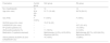
ResultsClinical features of examined groupsDifferent groups were compared in this study which includes healthy controls (HC, n=10), group of lupus patients (SLE, n=14), and patients with rheumatoid arthritis (RA, n=14). SLE groups was further divided on the basis of SLEDAI score and comorbidity with arthritis. The median age of the participants was comparable across all groups. Majority of participants were women which was explained by the high prevalence of autoimmunity among women. Number of the SLE patients were in the active stage with median SLEDAI score of 15. While, the RA patients were in both remission and active stage, differentiated on the basis of DAS28 score. All the patients among both groups were on their routine therapies that can affect the immune response. Information related to therapy and clinical characteristics are presented in Table 1.
Clinical characteristics of studied group.
| Parameters | Control group | SLE group | RA group |
|---|---|---|---|
| No. of participants | 10 | 14 | 14 |
| Age (min–max) | 30.4 (25–40) | 31.71 (19–46) | 38.79 (29–51) |
| Sex (F/M) | F (100%) | F (100%) | F (100%) |
| SLEDAI score (min–max) | – | 15.07 (6–23) | – |
| DAS28-ESR (min–max) | – | – | 4.37 (2.3–6.3) |
| RF positive patients (%) | – | – | 14 (100%) |
| ANA positive patients (%) | – | 11 (78.5%) | 2 (14.2%) |
| Medication (% patients received) | – | Methotrexate (14.2%), HCQ (50%), Wysolone (64.2%) | Methotrexate (85.7%), HCQ (64.2%), Wysolone (28.5%) |
| Active patients (SLEDAI ≥5) and DAS28-ESR ≥3.2 | – | 14 (100%) | 12 (86%) |
List including description and abbreviation for the analysed 40 cytokines is provided in supplementary data (Table 1) which can be retrieved for future studies related to metanalysis.
The significance values were separated from non-significant ones based on the −log10 p values of 2. The volcano plot was plotted between the log2 fold change and −log10 p values and significant one was marked with red.
Comparison of cytokines levels between the HC and SLE showed the significant changes among 15 cytokines out of 40 (Fig. 1). Most of these were decreased among SLE group which includes Gro-a/CXCL1, IL-9, TNF-b, RANTES/CCL5, MIP-1b/CCL4, IL-7, and MCP-3/CCL7 (Fig. 1A). Seven cytokines including SDF-1a/CXCL12, IL-18, IP-10/CXCL10, MIF, MIG, CSF and SCF were increased in SLE on comparing with HC group (Fig. 1B). Among those decreased in SLE, the Gro-a/CXCL1 was on the top with p value of 0.00001784 while the SDF-1a/CXCL12 on the top among increased one with p value of 0.0000965.
 Volcano plot showing log2 fold change vs −log p value pairwise comparison. (B) Bar graph showing log2 fold change in cytokines among healthy controls vs SLE patients.")
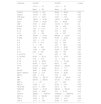
Assessment of cytokines levels based on the clinical parameters was performed where SLE patients were divided in two groups based on SLEDAI score. SLEDAI score of 12 considered to be point for distinguishing the low severe (n=6, <12 score) and high severe (n=8, >12 score) patients (Table 2). The level of significance was considered <0.05 during the comparison on SLEDAI basis. On evaluation among low severe and high severe patients, the concentration of cytokines such as MIP-1b/CCL4 (p=0.043), PDGF (p=0.041), IL-18 (p=0.042), IL-12 (p=0.02), and IL-9 (p=0.034) were found to be significantly increased in more severe lupus patients. These outcomes suggested the importance of cytokines in modulating the disease severity towards deprived state.
Comparison of cytokine levels in SLE patients based on SLEDAI score.
| Cytokines | SLEDAI | SLEDAI | p value |
|---|---|---|---|
| <12 (n=6) | >12 (n=8) | ||
| Mean±SD | Mean±SD | ||
| CTACK | 101.29±46.1 | 93.08±26.06 | 0.35 |
| Eotaxin | 15.016±9.92 | 11.70±8.04 | 0.26 |
| FGF basic | 19.11±16.07 | 16.97±4.79 | 0.38 |
| G-CSF | 148.87±97.26 | 106.57±68.08 | 0.19 |
| Gro-a | 116.48±26.44 | 59.5±56.28 | 0.25 |
| HGF | 72.47±35.44 | 151.65±53.87 | 0.2 |
| IFN-a2 | 10.75±8.44 | 7.28±3.56 | 0.21 |
| IFN-g | 7.30±11.13 | 7.19±11.74 | 0.49 |
| IL-1a | 4.93±6.33 | 3.73±4.54 | 0.35 |
| IL-1b | 1.33±1.54 | 1.18±0.57 | 0.41 |
| IL-1RA | 193.50±219.6 | 668.28±123.57 | 0.13 |
| IL-2RA | 79.53±119.95 | 55.34±32.33 | 0.32 |
| IL-4 | 1.43±1.12 | 1.12±0.32 | 0.27 |
| IL-6 | 5.75±5.14 | 6.07±5.40 | 0.47 |
| IL-7 | 3±1.53 | 3.57±1.82 | 0.26 |
| IL-8 | 7.49±4.67 | 21.57±26.81 | 0.09 |
| IL-9 | 10.66±5.89 | 32.46±25.85 | 0.03* |
| IL-10 | 6.53±8.31 | 3.62±1.56 | 0.21 |
| IL-12 (P70) | 1±0.54 | 2.35±1.34 | 0.02* |
| IL-12 (P40) | 62.45±56.14 | 53.17±30.30 | 0.36 |
| IL-13 | 0.79±0.18 | 1.0±0.32 | 0.07 |
| IL-16 | 51.55±45.36 | 72.71±101.69 | 0.30 |
| IL-18 | 36.39±22.19 | 68.21±8.55 | 0.04* |
| IP-10 | 214.76±161.97 | 260.49±209.32 | 0.32 |
| LIF | 35.82±46.58 | 25.14±11.1 | 0.30 |
| M-CSF | 48.87±72.96 | 35.17±16.56 | 0.33 |
| MCP-1 (MCAF) | 34.54±31.6 | 67.65±90.92 | 0.18 |
| MCP-3 | 2.08±0.12 | 2.03±0.58 | 0.43 |
| MIF | 720.81±355.72 | 1087.48±194.97 | 0.21 |
| MIG | 549.78±589.54 | 577.22±707.50 | 0.46 |
| MIP-1a | 2±1.57 | 2.71±3.65 | 0.31 |
| MIP-1b | 19.08±1.17 | 35.54±9.74 | 0.04* |
| PDGF-Bb | 22.10±8.55 | 44.57±4.5 | 0.04* |
| RANTES | 102.68±91.63 | 246.30±305.46 | 0.12 |
| SCF | 93.43±93.60 | 78.7±81.05 | 0.38 |
| SCGF-b | 28609.1±12767.87 | 41609.52±25093.45 | 0.11 |
| SDF-1a | 470.0±200.38 | 454.80±100.45 | 0.43 |
| TNF-a | 15.94±18 | 18.63±18.11 | 0.39 |
| TNF-b | 12.17±7.82 | 23.66±23.97 | 0.11 |
Comparison of cytokine profiles among HCs vs RA patients demonstrated the 18 differentially altered cytokines out of 40 with a significant cutoff of <2 of −log10 p value (Fig. 2). Most of these were increased in RA group which includes the MIP-1b/CCL4, PDGF, IL-9, IL-4, TNF-b, IP-10/CXCL10, RANTES/CCL5, SDF-1/CXCL12, HGF, MIP, TNF-a, and CTACK/CCL27 (Fig. 2A and B). The decreased levels of cytokines such as IL-15, G-CSF, MCP-3/CCL7, MCP-1/CCL2, IL-10, and Gro-a/CXCL1 was found in RA. Based on the p value, MIP-1b/CCL4 (p=0.000001) was highest among increase cytokines while IL-15 (p=0.000002) was lowest in RA patients when compared with the HC. No significant difference was observed during comparison on clinical basis (DAS28 score).
 Volcano plot showing log2 fold change vs −log p value pairwise comparison. (B) Bar graph showing log2 fold change in cytokines among healthy controls vs RA patients.")
On comparison with HC, both SLE and RA patients showed the six similar differentially altered cytokines (Fig. 3C). Out of which the SDF-1a/CXCL12, IP-10/CXCL10, MCP-3/CCL7 and Gro-a/CXCL1 were following the similar pattern in both diseases which can be utilised for distinguished the autoimmune patients from HC. Importantly, the level of cytokines such as IL-9, TNF-b, RANTES/CCL5 was noticeably high in RA patients. In contrast, the level of these cytokines was significantly decreased in lupus patients as observed on analysis of data from HC vs SLE. These outcomes describe the difference among both autoimmune disorders based on cytokines level which can be utilised to clinically differentiate the RA and SLE patients.
 Volcano plot showing log2 fold change vs −log p value value pairwise comparison. (B) Bar graph showing log2 fold change in cytokines among SLE vs RA patients. (C) Venn diagram showing the common significant cytokines among SLE and RA patients. (D) Venn diagram showing the common significantly altered cytokines among SLE with arthritis vs RA group.")
Cytokine profiling in SLE patients compared with RA patients. (A) Volcano plot showing log2 fold change vs −log p value value pairwise comparison. (B) Bar graph showing log2 fold change in cytokines among SLE vs RA patients. (C) Venn diagram showing the common significant cytokines among SLE and RA patients. (D) Venn diagram showing the common significantly altered cytokines among SLE with arthritis vs RA group.
To further differentiate between two autoimmune conditions, SLE and RA, the comparison between SLE vs RA demonstrated the changes among 13 cytokines out of 40 (Fig. 3A and B). The cytokines which were increased in RA were MIP-1b/CCL4, IL-9, PDGF, TNF-b, RANTES/CCL5, CTACK/CCL27, and Gro-a/CXCL1, while MIF, CSF, IL-18, M-CSF, IFN-a2, SCF were elevated in samples from SLE (Fig. 3A). Most of these cytokines followed the same pattern among respective diseased condition when compared with HC. In contrast, the level of Gro-a/CXCL1, RANTES/CCL5, TNF-b, which was found to be decreased in lupus during HC vs SLE comparison, was increased in RA on SLE vs RA analysis. However, it remains same in SLE i.e., decreased in HC vs SLE as well as in SLE vs RA, which suggest the difference among these two autoimmune disorders on the basis of cytokine profile. Similar to HC vs RA comparison, MIP-1b/CCL4 was also top in RA patients in SLE vs RA comparison, while IL-18 was on top in SLE vs RA instead of SDF-1a/CXCL12, which was highest in HC vs SLE. These outcomes pointed towards importance of cytokines level in diseased specificity as well as these can be utilized for distinguishing between RA and SLE pathology.
Incidence of arthritis in SLE patients is quite high therefore we have further divided the SLE patients in two groups: (a) with arthritis (n=5) and (b) without arthritis (n=9). On comparison between these groups, seven cytokines show the significant changes (p=<0.05) which includes eotaxin, IL-9, IL-12, RANTES/CCL5, SCF, TNF-a and b. Of these, four that were decreased in lupus arthritis group are eotaxin, IL-9, RANTES, and TNF-b. While the level of cytokines such as IL-12, SCF, and TNF-a was increased among lupus patients with arthritis. Interestingly, the levels of eotaxin with p value 0.00002 was lowest among those in patients with lupus and arthritis, however no such observation was made during the previous comparisons including RA/SLE vs HC or RA vs SLE. Pointedly, the level of IL-9, RANTES/CCL5, TNF-b was decreased in lupus patients with arthritis which was in contrast with RA where the level of these cytokines was increased. TNF-a emerged as an important cytokine which was increased in RA group among all the comparisons including lupus with arthritis which can be specific to RA as well as can be utilised to differentiate lupus patients on the basis of arthritis comorbidities (Fig. 3D).
Analysis of protein–protein interactions among the significantly changed cytokinesProtein interaction among significantly altered cytokine levels was determined by using string web tool enabling us to understand the functional relationship among different cytokines in various groups (Fig. 4). The confidence level was adjusted up to 0.7 (high confidence) and the analysis of chemokines in SLE showed multiple functional interactions between CXCL1, CXCL10, CCL5, CXCL12, and CXCL9, constituting a core complex (Fig. 4A). The interactions of this core complex with other cytokines/chemokines such as IL-10, IL-4, CSF3R, CCL7, CCL2, and CCL27 confirmed the possibility of synergic interplay among interleukins and chemokines in disease outcomes.
 HCs and SLE, (B) HCs and RA, (C) common among RA and SLE, and (D) SLE with/without arthritis. STRING web tool was accessed on 15 September 2023 for protein interaction analysis. Ligands for the interactions shown at the bottom.")
Protein–protein interaction networks among significantly changed cytokines of (A) HCs and SLE, (B) HCs and RA, (C) common among RA and SLE, and (D) SLE with/without arthritis. STRING web tool was accessed on 15 September 2023 for protein interaction analysis. Ligands for the interactions shown at the bottom.
Interactions among significant cytokines from HCs vs RA shows the multifactorial interactions among the CXCL10, CCL5, CXCL12, CXCL9, CXCL1, and CXCL27 (Fig. 4B). The interactions of this core complex with other cytokines such as IL-7, IL-18, CSF3, and CCL7 prompt us to assume the synergistic effect of these cytokines in RA pathology. The multifunctional interaction among CXCL1, CXCL10, CCL5, and CXCL9 was common among both HC vs SLE/RA which pointed towards the importance of this complex core in modulating the autoimmune diseases and can also act as biomarkers for its diagnosis.
Further evaluation of interaction among common cytokines in SLE vs RA group demonstrate the close functional interaction among CXCL10, CXCL12, CXCL1, and CCL5 which was also observed among HCs vs SLE/RA (Fig. 4C). This core complex can also interact with the IL-9 and CCL7 which can also be associated with presence of arthritis manifestations among lupus patients as IL-9 reflected resemblance with arthritis conditions.
Analysis of the significantly altered cytokines among lupus patients with arthritis and without arthritis for protein interaction shows strong functional relation between CCL5, CCL11, TNF-a and TNF-b (LTA) (Fig. 4D). The involvement of CCL5 was common in all the comparisons thereby demonstrating its potential role in arthritis manifestations.
Correlation network among cytokinesThe interactions among cytokines can alter the level of others. A correlation network can expose these alterations which can be important in autoimmune conditions. Correlation matrix was run and visulaized based on the Fruchterman–Reingold algorithm. Correlation was plotted with p value of <0.01 and cytokine correlation network in HC showed small clusters with very few cytokines correlating with each other (Fig. 5A). Most of the correlations were positive while a negative correlation emerged between IL-7∼M-CSF, IL-7∼SCGF-b, and SCGF-b∼G-CSF. While in SLE, a complex cluster was observed where very dense population of cytokines was correlated and shows the synergetic function of cytokines in lupus pathology (Fig. 6B). Inside this cluster LIF, FGF, and IP-10/CXCL10 appeared as major player involved in positive correlation with others while in HC, lesser connections among cytokines were observed, thereby reflecting their importance in modulating the disease pathology. In SLE, a negative correlation was observed among Gro-a∼FGF∼IP-10/CXCL10 and b-NGF∼IL-10, showing the similar pattern as observed during the comparison of HC vs SLE.
 HCs, (B) SLE, and (C) RA. The p value of <0.01 was considered significant for correlation. Each cytokine is represented by blue circle.")
 HCs, (B) SLE, and (C) RA. The p value of <0.01 was considered significant for correlation. Each cytokine is represented by blue circle.")
 HCs vs SLE, (B) HC vs RA, and (C) SLE without vs with arthritis. The ROC curve analysis was performed and AUC value of >0.5 was considered for predict the diagnostic biomarker. Prediction of candidate drug based on the DSigDB database. (D) Table presenting top 10 drug molecules. (E) Clustergram showing the drugs ranked based on the p value.")
 HCs vs SLE, (B) HC vs RA, and (C) SLE without vs with arthritis. The ROC curve analysis was performed and AUC value of >0.5 was considered for predict the diagnostic biomarker. Prediction of candidate drug based on the DSigDB database. (D) Table presenting top 10 drug molecules. (E) Clustergram showing the drugs ranked based on the p value.")
Prediction of significantly altered cytokines as diagnostic biomarker. (A) HCs vs SLE, (B) HC vs RA, and (C) SLE without vs with arthritis. The ROC curve analysis was performed and AUC value of >0.5 was considered for predict the diagnostic biomarker. Prediction of candidate drug based on the DSigDB database. (D) Table presenting top 10 drug molecules. (E) Clustergram showing the drugs ranked based on the p value.
Interestingly, correlations observed in RA were between very few cytokines in comparison with SLE (Fig. 5C). Three different clusters have been formed with positive correlations. IL-7∼G-CSF∼b-NGF and IL-12 make one cluster. IL-18 was involved in center of the two, where one cluster was formed with HGF, IL-13 and RANTES while the other included IL-9, SCF-b/CXCL12, MIF, TNF-b and M-CSF and MCP-1. Complexity was higher in SLE correlations confirming the systemic nature of disease while the correlation among RA demonstrated the importance of a few cytokine network in modulating the localised autoimmune pathology.
ROC curve for the significantly changed cytokinesTo determine the diagnostic effectiveness of the significantly changed cytokines level among different groups, ROC analysis was performed in which the AUC value of >/=0.9 was considered for an effective biomarker (Fig. 6). ROC analysis among HCs vs SLE demonstrates the possibilities of Gro-a/CXCL1, IL-7, IL-18, MIF, M-CSF, and SDF-1a/CXCL12 with diagnostic effectiveness (Fig. 6A). Among these, the Gro-a/CXCL1 shows the AUC of 1 demonstrating its strong potential as biomarker. In HCs vs RA group, the following cytokines demonstrated good biomarker effectiveness: IP-10/CXCL10, HGF, MIP-1a/CCL3, RANTES/CCL5, SDF-1a/CXCL12, TNF-a, TNF-b, IL-4, PDGF, IL-9, MIP-1b/CCL4, G-CSF, and Gro-a/CXCL1 (Fig. 6B) as they had AUC of 1 and can be utilized to distinguished the RA patients from the HCs.
SLE patients were distributed based on the comorbidities of arthritis and evaluated for diagnostic difference among with arthritis vs without arthritis condition. On ROC analysis, the cytokines such as eotaxin, TNF-a, and SCF emerged out as potent biomarkers to differentiate lupus patients among aforesaid group. On comparison among lupus with arthritis vs RA, we observed TNF-a, IL-9, and RANTES/CCL5 as common cytokines in both conditions with good AUC, pointing towards their application in diagnostics of arthritis condition among HCs as well as in lupus patients.
Candidate drug predictionPrediction of candidate drug was done by using Enricher platform based on the DSigDB database in which cytokines common among both the diseases vs HCs i.e., CXCL1, CCL5, CXCL10, CXCL12, CCL7, and IL-9 were uploaded (Fig. 6D and E). Top 10 predicted therapeutic compounds were selected based on the p value which can target most of the selected cytokines (Fig. 6D). Among these compounds, acetovanillone (CTD00002374), titanium dioxide (CTD00000489), and phencyclidine (CTD00005881) target majority of the cytokines and have least p value (Fig. 6E). Acetovanillone is a plant-based ketone which exhibits potent anti-inflammatory and anti-oxidants properties. Further investigations among implication of these drugs in autoimmune disorders will provide a novel therapeutic regime.
Discussion:The outcomes obtained pointed towards the importance of cytokines profiling in differentiating the SLE and RA patients from healthy controls. Many cytokines emerge out as potential biomarkers which can differentiate the disorder on the basis of severity and comorbidities. It is crucial to note that the all patients involved in this study were on the therapies which can affect the immune response including cytokines levels. Beside this, we have observed the significantly altered level of cytokines among SLE patients including the increased level of SDF-1a/CXCL12, IL-18, IP-10/CXCL10, MIF, M-CSF, SCF which was also in accordance with the literature.8 The decreased Gro-a/CXCL1 and IL-9 levels in SLE was in oppose to literature where the Gro-a and IL-9 were considered as potential marker for diseases activity. The decreased RANTES and MIP-1b level was also in contrast to literature where the levels of both was positively correlated with diseases severity in SLE.9 This decrease among pro-inflammatory cytokines in lupus patients might be due to their daily medication. Elevated IL-18 level among SLE patients known to activates the Th1 and NK cells which in return secrets the chemokines such as CXCL10, CXCL12, etc.10 Increased level of CXCL10 and CXCL12 can recruit the neutrophils and T cells respectively, which can initiate and assist in immunopathological mechanisms in lupus patients.11 Activation of immune cells by interleukins and recruitment of immune cells by chemokines leads to the formation of a pathogenic loop which can be one of the causes in lupus complexity. Elevation in MIF level was known to reverse the anti-inflammatory effect of glucocorticoids, while the increased in SCF level might enhance the proliferation of myeloid cells having importance in lupus pathology. Thus, the obtained data pointed towards the interplay of pro-inflammatory cytokine and chemokines which can modulates the SLE pathogenesis.
Interestingly, the levels of cytokines such as MIP-1b/CCL4, IL-9, 1L-18, IL-12 and PDGF was increased in more severe patients with SLEDAI score >12. CCL4 was demonstrated in recruitment of monocytes and T cells and IL-12 and IL-18 can induce the Th1 response which facilitates the type I response including secretion of IL-9.12 PDGF can recruit the mast cells and IL-9 triggers the secretions from mast cells to accelerate the immune response.13 Therefore, the interplay of elevated cytokines among severe patients shows the importance of type 1 immune response in exacerbating the lupus towards more severity.
Protein interaction analysis shows that the majority of significantly altered cytokines in HC vs SLE are functionally interconnected and multiple interaction among the CXCL1, CXCL10, CCL5, CXCL12, and CXCL9, constitute a core complex. Functional interconnection between core complex showed their close interplay in accelerating the immune response as CCL5 known to facilitates the macrophage-NK migration and T cell/DC interaction and CXCL1, CXCL9, CXCL10, and CXCL12 regulates the Th1 response, Th1, CD8, neutrophils and NK trafficking which can potentially modulates the immunobiology of SLE.14 Majority of these chemokines involved in T and B cell induced local and renal inflammation in lupus.15 Interaction of the core complex with the others cytokines such as IL-10, IL-4, CSF3R, CCL7, CCL2 and CCL27 demonstrates the induction of anti-inflammatory in response to inflammatory milieu through IL-4 and IL-10 secretion.16 However, no significantly difference was observed among IL-4 and IL-10 level on comparison among HC vs SLE group. CCL7 and CCL2 also contributes in local and renal inflammation in lupus nephritis.17 Taken together, these data indicate the impaired functional interactions among chemokines and interleukins enhanced the dysregulated immune response in SLE.
Correlation analysis among SLE showed the densely related cytokines when compared with healthy control's correlation matrix. LIF, FGF, and IP-10/CXCL10 were majorly involved in correlation as the central modulator in SLE. LIF and CXCL10 can potentially modulate the SLE manifestations including the lupus nephritis and was positively correlated with ANA level. Increased FGF level in lupus patients was found to be associated with neuropsychiatric lupus conditions. Negative correlation among Gro-a∼FGF∼IP-10/CXCL10 and b-NGF∼IL-10 demonstrated the response of anti-inflammatory cytokines such as IL-10 and b-NGF to elevated level of Gro-a/CXCL1 and CXCL10.18 ROC analysis showed the possibilities of these chemokines and cytokines (AUC value of >0.9) such as Gro-a/CXCL1, SDF-1a/CXCL12, M-CSF, MIF, and IL-7, IL-18 as potential biomarkers for SLE. However, further studies are required with more number of patients for better efficacy of these biomarkers.
Furthermore, the cytokine profiles in HC vs RA showed the increased level of various cytokines in RA patients such as the MIP-1b/CCL4, PDGF, IL-9, IL-4, TNF-b, IP-10/CXCL10, RANTES/CCL5, SDF-1/CXCL12, HGF, MIP, TNF-a, and CTACK/CCL27 and these observations were similar to the previous literature.19 Ability of CCL4 in recruiting the monocytes and T cells which can secrete TNF-a and IL-4, an important factor for B cell survival.20 But an impaired B cell response may also trigger the autoimmune response in RA which can exacerbates the disease condition. High plasma level of IL-9, CXCL10 and CXCL12 can also recruit the neutrophils at joints which can further tigger the inflammation by secreting NETs along with various inflammatory cytokines among RA patients.21 The cytokines that were observed to be lower among RA patients included IL-15, G-CSF, MCP-3/CCL7, MCP-1/CCL2, IL-10, and Gro-a/CXCL1. As majority of RA patients were on methotrexate dosing, that might be the reason for the reduced level of these chemokines.
PPI analysis among significantly altered cytokines from RA shows the core complex of the chemokines such as the CXCL10, CCL5, CXCL12, CXCL9, CXCL1 and CXCL27, which was similar to the SLE PPI analysis. Involvement of these chemokines as the central modulator among these two pathologies demonstrated its importance as potential biomarker as well as the targets for the various therapeutics. The interplay of these chemokines in RA pathology might be of importance as CXCL10/12, and CCL5 can recruit the immune cells at joints or in synovium of the RA patients and can also regulate the Th1 immune response in RA by facilitating the NETs secretions from the neutrophils.22 The interactions of the core complex with other cytokines such as IL-7, IL-18, CSF3, and CCL7 demonstrated their synergistic interplay as IL-7 is secreted from the inflamed tissue which can further activate the synovial fibroblasts to secret the GM-CSF, facilitating the recruitment of different granulocytes.23 IL-18 was demonstrated to be positively correlated with TNF-a and IL-1b level in synovial fluid which can harsh the disease condition.24
Correlation analysis among RA patients’ cytokines were plotted which showed the interaction among few cytokines when compared with SLE. IL-18, RANTES/CCL5, and G-CSF emerges out as the central connector. Majority of these were chemokines which can recruit the various immune cells contributing to the high inflammatory milieu in RA.25 Further analysis was performed to explore their importance as potential biomarker where the RANTES, TNF-b, IL-4, MIP-1b, G-CSF and IL-9 shows the AUC of 1. However, more extensive studies are required with large number of samples to confirm these findings.
SLE and RA involves the interplay of similar kind of cytokines which makes it difficult to distinguished between them and majority of the lupus patients experienced arthritis at least once during the active disease. Therefore, the comparison of cytokine profile was performed among both the diseases i.e., SLE vs RA, which shows the differentially altered cytokines from which majority of the cytokines were following the same pattern as of HC vs SLE/RA. The commonly altered cytokines among SLE and RA includes IL-9, Gro-a/CXCL1, RANTES/CCL5, IP-10, TNF-b, and MCP-3/CCL7. We have reported few common cytokines following the similar trends among both the pathologies such as SDF-1a/CXCL12 and IP-10/CXCL10, which were increased while MCP-3/CCL7 and Gro-a/CXCL1 were decreased. Rest of the common cytokines was considered to be specific for disease conditions as IL-9, RANETS/CCL5 and TNF-b was decreased among SLE patients while increased in RA. PPI interaction among common significantly altered cytokines in SLE vs RA shows a complex core which was similar to HC vs SLE/RA core which established the importance of CXCL1, CXCL10, and CCL5 amid both the pathologies. These cytokines have prominent AUC values therefore possess the potential to be utilized as biomarkers.
Further comparison was done among lupus patients on the basis of the presence or absence of arthritis. The significant alteration in cytokines shows the increased level of cytokines in patients with arthritis which includes IL-12, SCF, and TNF-a and decreased level of eotaxin/CCL11, IL-9, RANTES, and TNF-b. Generally, the level of all these cytokines is observed to be elevated among both the diseases,26 but the difference among with/without arthritis condition might be of clinical interest. Eotaxin/CCL11 can also be utilized as biomarker in lupus to discriminate between the non-nephritis vs nephritis patients.27 Moreover, a major fact remains that the alteration in the cytokines level can also be altered due to the prescribed treatment among the patients. The level of TNF-a was increased among the RA patients and lupus patients with arthritis while decreased in SLE patients including without arthritis conditions. This result shows the TNF-a as a potential indicator for arthritis condition. However, the level of TNF-a was also demonstrated high among the lupus patients.28 PPI interactions among common cytokines in lupus with arthritis vs without arthritis shows the multifunctional interactions of CCL5, CCL11, TNF-a and TNF-b (LTA). This core complex was different from all previous PPI interactions which might be specific for lupus with arthritis condition, however, CCL5 was observed as common player among all the PPI analysis and have good AUC value which further endorse its potential role among these disorders. CCL5 shows positive correlation with oxidative stress and can induce tissue damage in SLE and RA29 and was previously also proven as biomarker in RA which fans the possibility for CCL5 to be considered as a biomarker for autoimmune response.
The current scenario of therapy for most autoimmune condition includes therapeutics that target the symptomatic results of the disease and thus providing momentary relief but require a lifetime of medications and thorough management of the disease. Moreover, the autoimmune conditions have ability to induce other autoimmune conditions, thus we require some common targets which can be targeted in multiple manifestations. These potential biomarkers may be utilised to for prognostic purposes and can also act as futuristic therapeutic targets to alleviate the effects of disease pathogenesis in various autoimmune conditions such as SLE and RA. To further explore the possible therapeutics to target the potential cytokines that emerge during the different comparisons in the current study include CXCL1, CCL5, CXCL10, CXCL12, CCL7, and IL-9, top 10 predicted drugs were screened out from DSigDB database and among these drugs, acetovanillone (CTD00002374) observed to be a potential drug due its least p value and have targets for majority of aforesaid cytokines. Acetovanillone is a plant-based ketone which has been previously used to treat many disorders such as cardio and hepatotoxicity,30 however its role in targeting of SLE and RA is still undercover.
Limitations of any scientific study are important and help in building a better future study. The limitation of the current study is sample size; therefore, the obtained results should be inferred with caution. Another factor to be borne in mind is to remember that the patients are on treatment which might influence the cytokines levels. Although the cytokines interplay can be of similar nature among majority of autoimmune disorders, however, their implication as biomarkers requires more extensive studies. The outcomes of this study draw a conclusion where the differential interplay of cytokines varies among both disorders, SLE and RA, which can provide a diagnostic approach to differentiate among both the pathologies based on the cytokines level. In addition to this, the common cytokines which emerges out from various comparison pointed towards the similar mechanisms involved in both diseases which can be utilised as targets as well as prognostic paradigm for their detection. SLE patients can include arthritis as one of the important comorbidities which makes the disease more severe and these patients observed different cytokine alterations from non-arthritic ones, which can also assist in planning of the therapeutic regime. Hence, the present study gives important pointers towards the relevance of using various cytokine profiles as biomarkers for differentiating the two autoimmune disorders. At the same time, therapeutic targets based on differences between the two disorders can be closely followed for futuristic studies.
CRediT authorship contribution statementAA, RB, and JK contributed to analysing of the data, designing of the study, and writing the manuscript. AB and AS provided the critical contributions and reviewing the manuscript.
Human and animal ethical permissionsHuman ethical permission was approved by post graduate institute of medical education and research (PGIMER), Chandigarh (IEC# IEC-05/2016-419 and IEC-01/2022-2277).
FundingWe acknowledge the University Grant Commission (UGC) for fellowship to Mr. Akhil. Financial assistance received from Department of Biotechnology (DBT)-BUILDER project to carry out this work are also acknowledged.
Conflict of interestThe authors declare no conflict of interest.
The following are the supplementary data to this article:


