Musculoskeletal ultrasound is groundbreaking in making direct assessment of diseases with musculoskeletal symptoms because of its accessibility and usefulness in diagnosis and in the treatment of many diseases of the locomotor apparatus.1–3 Its use quickly resolves doubts on differential diagnosis and accelerates decision making.
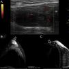
We recently assessed a woman aged 26, a new mother of 3 months who had presented at the emergency department on 3 occasions over 15 days due to right paracervical pain. The patient had no other history of interest. She took exercise sporadically and had an athletic constitution. On 3 occasions she had been diagnosed with mechanical cervicalgia associated with muscular contracture attributed to the tasks of looking after a newborn. She consulted for a fourth time after failure of 3 lines of analgesic, muscle relaxant and anti-inflammatory treatment. There was obvious asymmetry of the trapezius muscles with swelling of the right and limitation in rotation and passive lateralisation of the neck towards the left and lateralisation towards the right. An X-ray of the cervical spine made in the second assessment showed no changes to the axial vertebral disposition. Due to the striking lack of improvement an ultrasound scan of the right trapezius muscle was performed where a tumour was found to be contained within its thickness, with smooth and well defined edges, homogeneous in appearance, hypochoic with respect to the adjacent muscle tissue and with a positive power Doppler signal (Fig. 1A). The longitudinal diameter of the mass was 7cm and anterolateral diameter was 5.5cm.
(A) Trapezius (TP) ultrasound scan, longitudinal slice on cephalic edge. A hypoechoic mass is observed, homogenous in appearance and completed contained within the thickness of the muscle with no contact with the fascia (F) or subcutaneous cellular tissue (SCCT). The power Doppler signal is essentially concentrated in peripheral regions. (B) NMR of the shoulder, DP/SPIR sequence to remove fatty tissue. Transversal slice of the trapezius (arrow). In its interior a hyperintense structure with well defined edges is enhanced and this occupies almost all of the muscle matter (*). (C) NMR of the shoulder, T1 sequence, sagittal slice. In this projection the longitudinal extension of the trapezius (arrow) is appreciated and the occupation by the same fusiform, hypointense structure, with respect to the adjacent tissues. (*).
On suspicion of a tumour dependent on striated tissue the patient was admitted to hospital for further study. NMR imaging confirmed the finding of a tumour mass contained within the right trapezius muscle (Fig. 1B and C). After fine needle aspiration and complete resection a 158g specimen was extracted, the pathological analysis of which revealed low grade myofibroblastic sarcoma (LGSM) which did not affect the resection margins.
LGSM are atypical tumours which are rare and usually diagnosed in the neck and oral cavity.4,5 They are composed of an uncontrolled proliferation of myofibroblasts which are connective tissue cells that share physiological properties with smooth muscle cells and firbroblasts.6,7 These tumours have a high tendency to produce early metastasis and local relapses.6–8 Most of shoulder sarcomas usually present in the deltoid muscles. There have been no reported cases of myofibroblastic sarcomas of the trapezius in the literature, although it may be the exceptional location of other tumours.9
Signal capture from power Doppler is an unequivocal sign of hyperaemia which in the context of tumours occupying spaces denotes inflammatory (infectious or not) activity or neoangiogenesis (correlationable with neoprolipherative processes).10
Notwithstanding the prevalence of these sarcomas in the described location, we believe it is right to point out the importance ultrasound of the locomotor apparatus has in differential diagnosis of musculoskeletal processes with atypical disease courses and to highlight its use in identification of expansive soft tissue tumours, especially those which demonstrate hyperaemia through power Doppler signal detection.
Ethical DisclosuresProtection of people and animalsThe authors declare that for this research no experiments have been carried out on humans or animals.
Data confidentialityThe authors declare that they have adhered to the protocols of their centre of work on the publication of patient data.
Right to privacy and informed consentThe authors declare that no patient data appear in this article
Conflict of InterestsThe authors have no conflict of interests to declare.
Please cite this article as: Guillen Astete CA, Larena Grijalva C. Sarcoma miofibroblástico de trapecio. Reumatol Clin. 2019;15:e47–e48.


