

To develop evidence-based recommendations on the use of ultrasound (US) and magnetic resonance imaging in patients with spondyloarthritis, including psoriatic arthritis, and juvenile idiopathic arthritis.
MethodsRecommendations were generated following a nominal group technique. A panel of experts (15 rheumatologists and 3 radiologists) was established in the first panel meeting to define the scope and purpose of the consensus document, as well as chapters, potential recommendations and systematic literature reviews (we used and updated those from previous EULAR documents). A first draft of recommendations and text was generated. Then, an electronic Delphi process (2 rounds) was carried out. Recommendations were voted from 1 (total disagreement) to 10 (total agreement). We defined agreement if at least 70% of participants voted ≥7. The level of evidence and grade or recommendation was assessed using the Oxford Centre for Evidence Based Medicine levels of evidence. The full text was circulated and reviewed by the panel. The consensus was coordinated by an expert methodologist.
ResultsA total of 12 recommendations were proposed for each disease. They include, along with explanations of the validity of US and magnetic resonance imaging regarding inflammation and damage detection, diagnosis, prediction (structural damage progression, flare, treatment response, etc.), monitoring and the use of US guided injections/biopsies.
ConclusionsThese recommendations will help clinicians use US and magnetic resonance imaging in patients with spondyloarthritis and juvenile idiopathic arthritis.
Establecer recomendaciones, basadas en la evidencia, sobre el uso de la ecografía (US) y la resonancia magnética en pacientes con espondiloartritis, incluyendo la artritis psoriásica, y en la artritis idiopática juvenil.
MétodosLas recomendaciones se consensuaron mediante metodología basada en grupos nominales. Un grupo de expertos (15 reumatólogos y 3 radiólogos) definió el alcance, los usuarios, los apartados, las posibles recomendaciones y las revisiones sistemáticas a realizar (se utilizaron y actualizaron las revisiones de documentos de consenso de EULAR), y se asignaron tareas. Los expertos delimitaron los apartados y redactaron las recomendaciones. El nivel de evidencia y el grado de recomendación se establecieron utilizando el sistema del Centre for Evidence Based Medicine de Oxford, y el grado de acuerdo mediante Delphi a 2 rondas. Las recomendaciones se votaron según una escala de 1 (total desacuerdo) a 10 (total acuerdo), definiéndose el acuerdo como una puntuación≥7 por al menos el 70% de los participantes. El documento fue revisado por los expertos y el proyecto estuvo coordinado por un metodólogo experto.
ResultadosSe emitieron 12 recomendaciones sobre la validez de la US y la resonancia magnética para la detección de actividad y daño estructural, capacidad diagnóstica, predictora (de progresión de daño estructural, brote de la enfermedad, respuesta al tratamiento, etc.), utilidad en la evaluación y monitorización del tratamiento, y uso de la US como guía (para infiltraciones, biopsias, etc.) en pacientes con espondiloartritis y artritis idiopática juvenil.
ConclusionesSe presentan unas recomendaciones útiles para el manejo de la US y la resonancia magnética por los clínicos en pacientes con espondiloartritis y artritis idiopática juvenil.
Ultrasound (US) and magnetic resonance imaging (MRI) are of great utility in the daily clinical practice of the rheumatologist, both in the diagnostic process and in the therapeutic management of inflammatory diseases. The development of new drugs and the establishment of criteria for a close control of the inflammatory activity have brought about a considerable change in the utilization of these two techniques in the routine management of patients with rheumatic diseases. The reason for this change lies both in the their capacity to detect inflammation (with the possibility of intensifying treatment and avoiding or reducing irreversible structural damage) and the increasing incorporation of US and the greater accessibility of MRI studies on the part of rheumatology departments.
Ultrasound has a great advantage in the fact that it can be performed at the point of care. This enables the immediate comparison with clinical data and findings from other studies in cases of diagnostic suspicion or doubts. As a consequence, it is essential to facilitate programmed learning according to a competitive curriculum for US in rheumatology, to gain access to a medium or high-range US machine and to become familiar with the settings. Magnetic resonance imaging may not be as accessible in the rheumatology department as US, but it is a highly useful imaging technique, both for the diagnosis and for patient follow-up.
The incorporation of these imaging techniques into clinical practice should be based on valid scientific criteria, judgment and feasibility. Therefore, the main objective of this project is to draw up recommendations on the use of US and MRI in spondyloarthritis (SpA), including psoriatic arthritis (PsA), and in juvenile idiopathic arthritis (JIA), based on the best available evidence, which serves as a reference for all of the professionals involved in caring for patients with rheumatic diseases. Our proposal is to reduce the variability in the use of these imaging techniques and to close gaps between clinical practice and the best scientific evidence.
Material and MethodsThe preparation of this document was an initiative of the Working Group on Ultrasound of the Spanish Society of Rheumatology (SER). The present article is to provide recommendations concerning the use of US and MRI in patients with SpA, including PsA, and in JIA patients. The recommendations on the use of these imaging techniques in rheumatoid arthritis are dealt with in another document. The development involved the utilization of the Delphi method and the nominal group technique. The entire process of writing the document was performed by distributing tasks and comments among those participating, with the additional aid of several consensus documents published by the European League Against Rheumatism (EULAR) and the critical evaluation and the subsequent update of their systematic literature reviews (SLR).1–3
Selection of the Panel and Assignment of TasksThe first step was the formation of a panel of 18 experts (15 rheumatologists and 3 radiologists), selected through a search in MEDLINE for Spanish professionals with publications in indexed journals on the utilization of US and/or MRI in SpA and JIA. The panel was constituted on the basis of the results of that search, the demonstrated experience of the professionals and their interest in the subject, also taking into account criteria of geographic representation. The entire process was coordinated by a methodologist with demonstrated experience in the Delphi method and SLR.
In the first meeting of the nominal group, the clinical questions to be developed were selected and the scope, objectives and sections of the document were decided. The clinical questions were formulated following the PICO format: patient, intervention, comparison and outcome. It was finally decided to carry out the SLR on different aspects of US and MRI in SpA and JIA, and postpone the assignment of tasks to the panelists until the results of the SLR had been obtained. Given that these clinical questions had been previously formulated in the abovementioned EULAR consensus documents, it was decided to evaluate them critically and update them.
Systematic Literature ReviewsThe critical evaluation and updating of the SLR were performed with the help of an expert Spanish documentalist. For this, we contacted those responsible for carrying out the SLR of the consensus documents published by EULAR on SpA and JIA, to evaluate the questions and search strategies.1,3 The latter included the use of MeSH terms and free text such as “ultrasonography” [MeSH] or ultrasono* [tw]. The evidence tables and conclusions were also critically evaluated. In the SLR of these EULAR documents, we screened for the following bibliographic databases: MEDLINE (from inception to January 2013 and November 2013 for SpA and JIA, respectively) and Cochrane Library (from inception to January 2013 and November 2013 for SpA and JIA, respectively). The present document was updated from those dates to December 2014. Subsequently, using clinical queries, the bibliography was updated to May 2015. The search strategies of the EULAR documents were constructed combining terms in MeSH-like subject headings and free text, in order to improve and achieve a balance between the sensitivity and specificity. The expert documentalist evaluated them and considered them suitable, but introduced certain new terms to improve their yield. The inclusion and exclusion criteria were those used for the EULAR document. Regardless of the evidence tables of the EULAR documents, which were considered suitable, we began with the selection of articles with the search completed (that corresponding to the EULAR document and that of the update). The process of selecting the articles (using a reference manager) was done by 2 independent reviewers (the SLR were distributed by pairs, for a total of 21, 10 for SpA and 11 for JIA, for 2 reviewers, EL and MGY), who also analyzed in detail the articles retrieved with the search strategies, utilizing a data collection form designed for that purpose. The methodological quality of the studies included was evaluated using the Quality Assessment of Diagnostic Accuracy Studies (QUADAS) tool4 and Centre for Evidence-Based Medicine of Oxford,5 and a series of questions to evaluate the risk of biases and the applicability of the studies. In the end, we analyzed the level of evidence (LE) of each of the studies using the Oxford criteria because of the large volume of studies and the heterogeneity of the aspects evaluated (diagnosis, monitoring, etc.). The results of the SLR were also employed to establish the grade of recommendation (GR). All of the information in the studies was extracted from evidence tables. This entire process was supervised by an expert methodologist and 2 rheumatologists who were expert in the use of these imaging techniques.
Delphi StudyThe different sections of the document were distributed among the members of the panel who were to draw them up and for the preparation of the corresponding recommendation(s). They received a report of the results of the corresponding SLR to provide support for their drafts. Once drawn up and edited, the recommendations underwent the evaluation of the levels of agreement (LA) by means of a Delphi survey. For this, we sent the panelists an online questionnaire (http://www.surveymonkey.com) with the complete recommendations, together with the necessary instructions for voting by sending their LA for each of them (first Delph round). The LA was assessed by means of voting in a Likert scale from 1 (totally in disagreement) to 10 (totally in agreement), and agreement was defined by a score ≥7 voted by at least 70% of the participants. The overall results of the Delphi were sent to all of the panelists (modified Delphi). The recommendations with a LA of less than 70% were reedited and voted in a second round, in pertinent cases. In the first round, it was also possible to include new recommendations to eventually be voted in the second round.
Edition of the Final DocumentOnce the Delphi study was completed, the sections and recommendations were integrated and edited. The complete document was then sent to the group of panelists for the introduction of the corrections and the necessary comments, which resulted in a final report for the preparation of the definitive document. The methodologist participated in the assignment, of each of the recommendations, the LE and the GR according to the Centre for Evidence-Based Medicine of Oxford,5 and the LA (from Delphi). Once the process was concluded, everything was sent to 2 external reviewers, a clinical rheumatologist and a medical epidemiologist with extensive experience in the validation of imaging techniques. The systematic reviews will soon be submitted for publication to both international and Spanish journals. The process and final document were reviewed and endorsed by the SER.
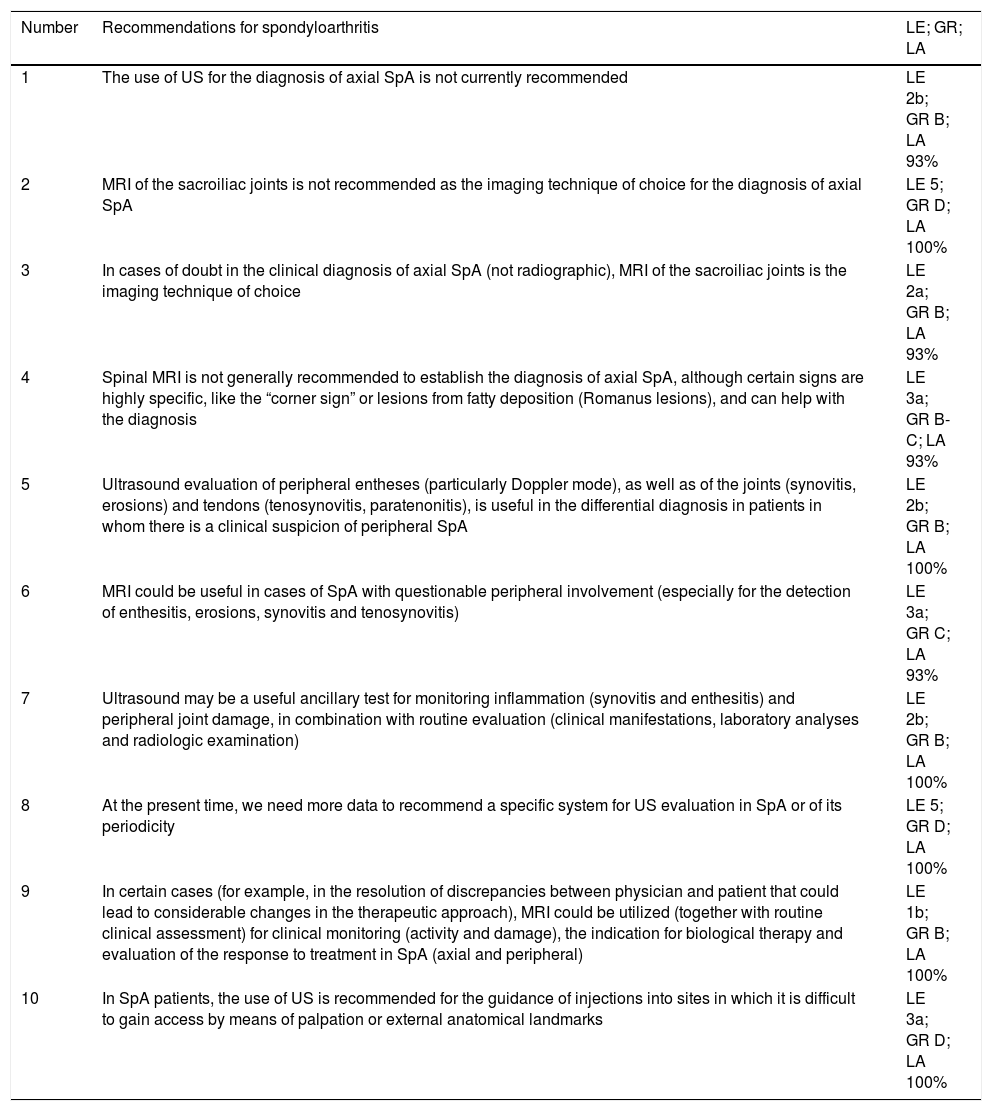
ResultsThe recommendations generated, which appear in Table 1, are described below.
Definition of the Recommendations, Together With the Level of Evidence, Grade of Recommendation and Level of Agreement, for Spondyloarthritis and Juvenile Idiopathic Arthritis, in Relation to Ultrasound and Magnetic Resonance Imaging.
| Number | Recommendations for spondyloarthritis | LE; GR; LA |
|---|---|---|
| 1 | The use of US for the diagnosis of axial SpA is not currently recommended | LE 2b; GR B; LA 93% |
| 2 | MRI of the sacroiliac joints is not recommended as the imaging technique of choice for the diagnosis of axial SpA | LE 5; GR D; LA 100% |
| 3 | In cases of doubt in the clinical diagnosis of axial SpA (not radiographic), MRI of the sacroiliac joints is the imaging technique of choice | LE 2a; GR B; LA 93% |
| 4 | Spinal MRI is not generally recommended to establish the diagnosis of axial SpA, although certain signs are highly specific, like the “corner sign” or lesions from fatty deposition (Romanus lesions), and can help with the diagnosis | LE 3a; GR B-C; LA 93% |
| 5 | Ultrasound evaluation of peripheral entheses (particularly Doppler mode), as well as of the joints (synovitis, erosions) and tendons (tenosynovitis, paratenonitis), is useful in the differential diagnosis in patients in whom there is a clinical suspicion of peripheral SpA | LE 2b; GR B; LA 100% |
| 6 | MRI could be useful in cases of SpA with questionable peripheral involvement (especially for the detection of enthesitis, erosions, synovitis and tenosynovitis) | LE 3a; GR C; LA 93% |
| 7 | Ultrasound may be a useful ancillary test for monitoring inflammation (synovitis and enthesitis) and peripheral joint damage, in combination with routine evaluation (clinical manifestations, laboratory analyses and radiologic examination) | LE 2b; GR B; LA 100% |
| 8 | At the present time, we need more data to recommend a specific system for US evaluation in SpA or of its periodicity | LE 5; GR D; LA 100% |
| 9 | In certain cases (for example, in the resolution of discrepancies between physician and patient that could lead to considerable changes in the therapeutic approach), MRI could be utilized (together with routine clinical assessment) for clinical monitoring (activity and damage), the indication for biological therapy and evaluation of the response to treatment in SpA (axial and peripheral) | LE 1b; GR B; LA 100% |
| 10 | In SpA patients, the use of US is recommended for the guidance of injections into sites in which it is difficult to gain access by means of palpation or external anatomical landmarks | LE 3a; GR D; LA 100% |
| Number | Recommendations for juvenile idiopathic arthritis | LE; GR; LA |
|---|---|---|
| 11 | US and MRI (synovial proliferation and joint effusion) can be utilized as ancillary tests for clinical evaluation in the diagnosis of JIA in children | LE 2a; GR B; LA 100% |
| 12 | Ultrasound is the technique of choice for image-guided injection in JIA | LE 2a; GR B; LA 100% |
GR, grade of recommendation; JIA, juvenile idiopathic arthritis; LA, level of agreement; LE, level of evidence; MRI, magnetic resonance imaging; SpA, spondyloarthritis; US, ultrasound.
Ultrasound is capable of detecting synovitis, tenosynovitis, paratenonitis, enthesitis and inflammation of connective tissue in patients with SpA. In terms of inflammation, MRI can also evaluate subchondral bone marrow edema (BME).
In the case of peripheral involvement, US and MRI may be useful for assessing inflammation, but always as ancillary tests for routine clinical evaluation.4–11 Nevertheless, a greater number of studies would be necessary to determine their role more accurately.
In cases of axial involvement and, again, as an ancillary test for clinical evaluation, MRI may be a useful imaging technique in certain cases. It has established the existence of BME at the level of the sacroiliac joints and spine in distinct forms of SpA, and it has also demonstrated the existence of an association between edema adjacent to the sacroiliac joint and the presence of inflammation revealed by histological examination.12
Value of Imaging Studies in the Evaluation of Structural Damage in SpondyloarthritidesBoth US and MRI could be useful in assessing chronic damage in SpA, again as an ancillary test for routine clinical evaluation.
Ultrasound is capable of detecting damage and structural changes in cases of peripheral involvement, such as erosions, calcifications in the entheses or within the tendons, especially in PsA patients.13 With respect to MRI, it has been found to be a sensitive technique for detecting bone erosions, especially in patients with rheumatoid arthritis.14,15
As in the case of inflammation, a greater amount of evidence is needed to issue more accurate recommendations on the role of these imaging techniques in the assessment of structural damage in SpA.
Diagnostic ValueRecommendation 1. At the present time, the use of US is not recommended for the diagnosis of axial SpA (LE 2b; GR B; LA 93%).
A number of data have been reported on the capacity of gray-scale US to detect sacroiliitis in SpA, based on the existence of effusion or changes in the sacroiliac ligaments.16,17 On the other hand, when compared with MRI, the use of Doppler US has shown high specificity for the detection of sacroiliitis in comparison with MRI, although with variable sensitivity, depending on the study.18,19 The use of intravenous contrast media improves the yield of US for the diagnosis of sacroiliitis, although its complexity limits its use in clinical practice.18–21 A recent study involving patients with inflammatory low back pain defined a series of ultrasound parameters with a diagnostic utility for sacroiliitis similar to that of MRI.22 However, these results should be studied in greater depth.
Recommendation 2. Magnetic resonance imaging is not recommended for the sacroiliac joints as the imaging technique of choice for the diagnosis of axial SpA (LE 5; GR D; LA 100%).
At the present time, plain radiography of the sacroiliac joints continues to be the most widely employed diagnostic imaging technique in these patients.
Recommendation 3. In cases of doubt concerning the clinical diagnosis of axial SpA (not radiographic), MRI of the sacroiliac joints is the imaging technique of choice (LE 2a; GR B; LA 93%).
The results of the different studies on the value of MRI of the sacroiliac joints for the diagnosis of SpA have had variable validity: sensitivity of 0.11%–0.98% and specificity of 0.43%–0.99% in patients with inflammatory low back pain or suspected axial SpA.23–29 This variability depends on the type of study, the study population and the criteria utilized to define inflammatory low back pain and axial SpA. We obtained moderate to high sensitivity and specificity values (0.82%–0.90% and 0.94%–0.97%, respectively) in established SpA,25,26,29,30 regardless of the diagnostic and/or classification criteria employed. Likewise, MRI has been found to produce better results than plain radiography or computed tomography for the diagnosis of sacroiliitis in patients with inflammatory low back pain (defined according to different criteria).31,32
Recommendation 4. Magnetic resonance imaging of the spine is not generally recommended to establish the diagnosis of axial SpA, although there are signs that are highly specific for axial SpA, like the “corner sign” or lesions from fatty deposition (Romanus lesions), and can help with the diagnosis (LE 3a; GR B-C; LA 93%).
Magnetic resonance imaging of the spine has been found to have a low-to-moderate sensitivity (0.44%–0.69%) for the diagnosis of axial SpA, with a variable specificity (0.43%–0.99%).24,30,33 Sensitivity for ankylosing spondylitis (AS) (New York criteria) is also low-to-moderate (0.44%–0.69%), although with a very high specificity (0.96%–0.97%).24,30,33,34 The results of one study have shown that the addition of MRI of the spine to that of the sacroiliac joint adds little to the value of early SpA detection and classification by MRI of only the sacroiliac joints.35
Recommendation 5. Ultrasound evaluation of peripheral entheses (particularly Doppler mode), as well as of the joints (synovitis, erosions) and tendons (tenosynovitis, paratenonitis), is useful in the differential diagnosis in patients in whom there is a clinical suspicion of peripheral SpA (LE 2b; GR B; LA 100%).
When enthesitis is found by US, the findings are capable of discriminating between patients with PsA or SpA and healthy individuals and those with fibromyalgia.36,37 Moreover, US is more sensitive and specific than physical examination for detecting entheseal involvement in patients with SpA,38–40 established AS41 and PsA.36,42
With regard to the diagnosis of peripheral SpA, on comparing the clinical diagnostic criteria, Doppler mode for enthesis was moderately sensitive (0.76%) and had a high specificity (0.81%) in patients with suspected peripheral SpA.36 On the other hand, its sensitivity was low and specificity high in early SpA, and it had an elevated sensitivity (0.83%–0.98%) and a variable specificity (0.48%–0.9%) in established peripheral SpA,36,39,43 and a low-to-moderate sensitivity (0.36%–0.72%) and a moderate specificity (0.67%–0.76%) in patients with PsA.36,37,44,45
The predictive value of US using multivariate analysis was assessed in only 1 study. The results confirmed that US was an independent predictor of the diagnosis of peripheral SpA.46 Ultimately, the detection of enthesitis with Doppler mode was associated with an early diagnosis of SpA, even if only 1 enthesis was involved.45 There are several enthesitis indices that can contribute to reaching a diagnosis of SpA and early SpA, but, at this time, it is not possible to recommend any specific one, or a particular enthesitis score or count.38,41,43,47
Recommendation 6. Magnetic resonance imaging may be useful in cases of SpA with questionable peripheral involvement (especially for the detection of enthesitis, erosions, synovitis and tenosynovitis) (LE 3a; GR C; LA 93%).
The results of one study have demonstrated that MRI of the heel has a very high specificity (0.94%), although a low sensitivity (0.22%), for distinguishing patients with SpA (according to the Amor criteria) from controls (noninflammatory degenerative disease).48 Further studies are needed to determine the diagnostic value of MRI in peripheral SpA.
Prognostic ValueIn methodological terms, it is very important to take into account that research on the course of these diseases requires the conduction of longer-term longitudinal studies than those focusing on rheumatoid arthritis, especially in cases of early SpA. Thus, the results shown below on the predictive value of these imaging techniques should be interpreted with caution due to the shorter duration of the studies.
At the present time, we do not have sufficient evidence on the predictive value (of pain, remission or response to treatment) of US in SpA.49,50
On the other hand, in some longitudinal studies, an association has been observed between the presence of baseline inflammation defined by BME (in MRI of the sacroiliac joints and spine with and without contrast) and the development of chronic changes in sacroiliac joints, detected both in MRI and in plain radiography. Baseline BME has also been related to the development over time of subchondral fatty deposition and erosions.51
Likewise, it has been observed that the reduction of sacroiliac inflammation (after treatment with anti-tumor necrosis factor-α [anti-TNF-α] agents), revealed by MRI, is associated with the formation of syndesmophytes at the vertebral level according to plain radiography.52 Moreover, it is known that there is a relationship between the resolution of the inflammation shown by MRI and the development of fatty degeneration in the spine and the sacroiliac joints.52–57 After treatment, BME in the sacroiliac joints and spine resolves, but fatty deposition on subchondral bone is more frequently associated with the development of syndesmophytes on the spine.
The presence of baseline BME in MRI of the knee does not appear to have any predictive value concerning structural changes in plain radiography or important changes in quality of life at 10 years.58
RemissionThe available information on the value of US and MRI in SpA patients in remission is scarce at the present time.
Evaluation of and Monitoring the Therapeutic ResponseRecommendation 7. Ultrasound may be a useful ancillary test for monitoring inflammation (synovitis and enthesitis) and peripheral joint damage in combination with routine evaluation (clinical manifestations, laboratory analyses and radiologic examination) (LE 2b; GR B; LA 100%).
Recommendation 8. At the present time, we need more data to recommend a specific system for US evaluation in SpA or of its periodicity (LE 5; GR D; LA 100%).
We have no evidence of the possible role of US in clinical monitoring (in terms of activity, damage and response to treatment) of axial involvement in SpA.
The existence of a possible association between US parameters and clinical and/or laboratory evidence of activity has been observed in certain cross-sectional studies but not in others.39,59–63 On the other hand, the results of a prospective study showed that a number of US findings in elemental lesions of the enthesis may be sensitive to change in response to an effective treatment.64
On the other hand, there are several enthesitis indices developed for the diagnosis of SpA that have not been utilized for monitoring that condition.
Recommendation 9. In certain cases (for example, in the resolution of discrepancies between physician and patient that could lead to considerable changes in the therapeutic approach), MRI could be utilized (together with routine clinical assessment) for clinical monitoring (activity and damage), the indication for biological therapy and evaluation of the response to treatment in SpA (axial and peripheral) (LE 1b; GR B; LA 100%).
Magnetic resonance imaging of sacroiliac joints and lumbar spine has been found to be sensitive to the change in SpA (in patients classified according to different criteria, and being treated with distinct biological agents).65–70
No association has been observed between baseline inflammation (or its changes) in MRI of the sacroiliac joints and the Bath Ankylosing Spondylitis Disease Activity Index – BASDAI,27,71,72 C-reactive protein,66,73 pain or other activity variables.27,66,72–75 The results referred to in the Ankylosing Spondylitis Disease Activity Score – ASDAS – are insufficient.65,72 With respect to spinal MRI, most of the studies analyzed show no relationship between inflammation (including BME) and the BASDAI,66–68,71,72,76–79 the ASAS 20 of the Assessment in Ankylosing Spondylitis Working Group,78 pain,66,78 or peripheral enthesitis.77 Data related to C-reactive protein or erythrocyte sedimentation rate are contradictory.66–68,72,77–80 However, a relationship to changes in the ASDAS has been observed in several studies.65,72,78
With respect to monitoring structural damage in patients with SpA, a relationship has been demonstrated between structural damage in MRI of the spine and sacroiliac joint (measured in different sequences by predefined scores of certain specific findings, such as fatty marrow depositions) and the progression of radiographic damage,51,81–83 although the data are contradictory with respect to structural changes detected by computed tomography.74,83 Until now, no study has been able to demonstrate the existence of a firm relationship between spinal MRI and the Bath Ankylosing Spondylitis Functional Index, clinical examination or the Schober test.74,83 However, an association between structural damage and MRI and lumbar lateral flexion has been reported.84,85
In the presence of peripheral involvement, no significant correlations have been observed between BME in hands or knees and other clinical variables of activity, such as the Disease Activity Score in 28 Joints or C-reactive proteins, that justify the use of MRI to monitor the activity of SpA.58,69,86 With respect to monitoring structural damage, there are preliminary data of a cross-sectional study that reveal a relationship between BME and the erosions detected by MRI and plain radiography of the hands.86
Finally, the available data is too scarce to support the recommendation of the frequency and type of monitoring of the inflammatory activity and axial and peripheral structural changes in SpA using MRI.87–92
Predicting the Response to TreatmentIn the case of US, additional studies will be necessary to establish the predictive value of the response to treatment in SpA. The available data concerning MRI are also insufficient. It has been observed that, in patients with SpA being treated with anti-TNF-α, spinal MRI can predict the BASDAI 50 score at 3 months.93 In another study, baseline inflammation in the spine and sacroiliac joints was associated with a better response to adalimumab at 12 weeks.75
Guided InjectionRecommendation 10. In SpA patients, the use of US is recommended for the guidance of injections into sites in which it is difficult to gain access by means of palpation or external anatomical landmarks (LE 3a; GR D; LA 100%).
At the present time, there is not enough evidence that guided injection is an improvement on routine injection in patients with SpA.94–98 However, the panel, on the basis of experience, considered that it may be useful in these cases.
Juvenile Idiopathic ArthritisRecommendation 11. Ultrasound and MRI (synovial proliferation and joint effusion) can be utilized as ancillary tests for clinical evaluation in the diagnosis of juvenile idiopathic arthritis (JIA) in children (LE 2a; GR B; LA 100%).
Recommendation 12. Ultrasound is the technique of choice for image-guided injection in JIA (LE 2a; GR B; LA 100%).
At this time, as both US and MRI are considered valuable ancillary tests in JIA, the choice of one or the other will depend on the joint or joints to be examined and the objective of the indication. The need to sedate the child and the availability of the technique in the routine practice of the department or center will be other factors to be considered.
The published literature demonstrates that US and MRI are more sensitive for the detection of inflammation in children than physical examination and plain radiography. Moreover, they enable the detection of synovitis and the establishment of the differential diagnosis with respect to periarticular processes.99,100
There is evidence of a certain correlation between clinical activity (including analytical parameters) and the 2 imaging techniques (US and MRI). Although this correlation appears to increase with the inclusion of Doppler in the US study and intravenous contrast in MRI, to date, there are few studies supporting these findings.101,102
Because of the particular features of the growing skeleton, the detection of structural damage in children with JIA is one of the major challenges to be considered in the utilization of these imaging studies. At the present time, the available evidence is too limited to determine whether these imaging techniques enable the differentiation of whether the changes on the joint surface actually reflect structural damage or are merely part of normal development.103
With respect to the diagnosis, at this time, there are no specific US or MRI findings that make it possible to establish a definitive diagnosis for JIA. The medical records—supported by the ancillary tests—continue to be the cornerstone in this process.
In contrast to MRI, US enables the examination of multiple joints and the determination of the extension of JIA and, thus, the early classification of the children in the different categories. To date, just one study has focused on determining the minimum number of joints that must be included in the evaluation of the extension of joint involvement.104 At least the following joints should be included: knees, ankles, wrists and second metacarpophalangeal joints.
Despite the evidence accumulated concerning these 2 imaging tests in terms of their utility in the prognosis and monitoring of the therapeutic response in rheumatoid arthritis, research performed in these settings is nearly nonexistent in the case of JIA.105
Preliminary data from a 1-year follow-up with contrast-enhanced MRI indicate that the automatic quantification of the baseline inflamed synovial membrane volume could help to detect progression of erosion.106
Most of the studies performed to assess monitoring of the therapeutic response in JIA are based on the local effect obtained with intra-articular injections. There are few reports that measure the response to drugs using imaging techniques.105
One advantage of US over MRI lies in its utilization for the guidance of injections. The results of 2 studies have shown that the course was more favorable in children with JIA who were treated with image-guided injections.107,108
The data on the few studies conducted to date in children with JIA in clinical remission do not enable us to define possible variations in the therapeutic approach based only on changes in the imaging techniques.
Finally, ultrasound is the imaging technique of choice because of its feasibility, accessibility and safety, when compared with other techniques, for the periodical evaluation of children with JIA.
DiscussionThese are the first recommendations that count on the participation of the SER. They endorse the use of US and MRI in clinical practice in patients with SpA and JIA, basing their support on the best evidence currently available.
Ultrasound and MRI were introduced into clinical practice and clinical trials as ancillary tests in the clinical evaluation of SpA and JIA, especially in the case of US, the performance of which is in the hands of the clinicians themselves in a growing number of centers in Spain. The most important added value of the two techniques is their greater sensitivity for the detection of synovitis, enthesitis and structural damage than standard physical examination and plain radiography. In recent years, publications on the metric properties of these techniques (validity, reliability and sensitivity to change) and on their diagnostic and predictive value have proliferated. However, there has yet to be a definition of or a consensus on their use in rheumatology clinical practice.
Therefore, for the purpose of improving clinical practice, we must establish explicit recommendations that encompass aspects as important as the diagnosis or monitoring of treatment. Although it is certain that the evidence is insufficient in certain areas, this document presents a series of highly relevant recommendations that can be especially useful for clinicians. Moreover, for the implementation of the entire project, we utilized several consensus documents published by EULAR and from the critical evaluation and resulting update of their SLR,1–3 as reference materials. These sources, together with all the methodological work carried out, lend great validity to this document.
Ethical DisclosuresProtection of human and animal subjectsThe authors declare that no experiments were performed on humans or animals for this investigation.
Confidentiality of dataThe authors declare that no patient data appears in this article.
Right to privacy and informed consentThe authors declare that no patient data appears in this article.
FundingFinanced by the Extraordinary Professorship accorded by the Universidad Complutense de Madrid and Merck Sharp & Dohme (UCM/MSD): Prof. Luis Carreño in Autoimmune Inflammatory Diseases.
Conflict of InterestEsperanza Naredo has received fees for presentations from Abbvie, Roche Farma, Bristol-Myers Squibb, Pfizer, UCB and Novartis.
Estíbaliz Loza has received fees for research projects from Abbvie, Roche Farma, Bristol-Myers Squibb, Pfizer, MSD, UCB, Sanofi-Aventis and Novartis.
Paz Collado has received fees for presentations from Abbvie and Pfizer.
Enrique Batlle has received fees for presentations, courses and/or projects and/or has worked as a consultant for Abbvie, BMS, Lilly, Menarini, MSD, Pfizer, Roche, UCB, Menarini and the Spanish Foundation of Rheumatology (FER). Victoria Navarro-Compán has received fees for presentations and for research projects from Abbvie, BMS, MSD, Novartis, Pfizer, Roche, UCB, SER and ASAS group.
Esther Vicente has received fees for presentations from Abbvie, Roche Farma, Bristol-Myers Squibb, Pfizer, UCB, ROVI and MSD.
Pilar Macarrón has received fees for presentations from Abbvie, Roche Farma, Bristol-Myers Squibb, Pfizer, MSD and UCB.
Carlos Acebes has received fees for presentations from Tedec-Meiji Farma.
The remaining authors declare they have no conflicts of interest.
This report is supported by the Spanish Society of Rheumatology (SER).
Please cite this article as: Uson J, Loza E, Möller I, Acebes C, Andreu JL, Batlle E, et al. Recomendaciones para el uso de la ecografía y la resonancia magnética en pacientes con espondiloartritis, incluyendo la artritis psoriásica, y en pacientes con artritis idiopática juvenil. Reumatol Clin. 2018;14:27–35.


