


We present the case of a 39-year-old woman with an egg-shaped tumor on the lateral aspect of her right ankle below the lateral malleolus, measuring 3.2cm. The lesion was detected by the patient 3 years before she came to the clinic; however, it produced pain when walking only a year before her visit. The tumor was painful on palpation and had a hard consistency (Fig. 1A). An ultrasound with Esaote MyLab 25® equipment and a multifrequency linear transducer of 10–18MHz, was used; we found an oval image with anechoic areas within it alternating with isoechoic regions (Fig. 1B). The lesion could not be compressed with the transducer and, with the application of power Doppler, vascularity was positive (Fig. 1C). The patient underwent surgery with complete removal of the lesion without complications. The histological study showed a benign mesenchymal neoplasm composed of smooth muscle bundles arranged in a disorganized way, with extensive areas of hyalinization and vascular thickening, changes consistent with the diagnosis of leiomyoma (Fig. 1D).
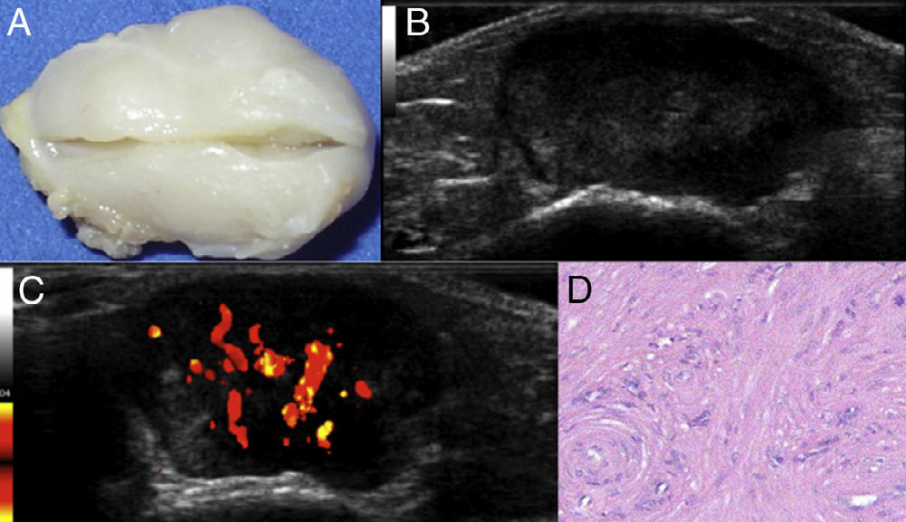
(A) Specimen obtained by surgical resection. (B) Grayscale ultrasound of the longitudinal axis of the lesion, showing defined edges, with alternating anechoic and isoechoic areas. (C) Positivity of the power Doppler signal (vascularity). (D) Histological section (hematoxylin–eosin) showing disorganized bundles of spindle cells (smooth muscle) and vascular thickening.
Leiomyomas arising outside of the uterus and the gastrointestinal tract are rare. These benign neoplasms of smooth muscle have been reported mainly in the lower limbs and feet of women between the third and fifth decade of life.1 The angioleiomyoma or vascular leiomyoma originates from the tunica media of small veins or arteries.2,3 Leiomyomas were divided into 3 groups: cutaneous, soft tissue (angioleiomyomas) or deep retroperitoneal or on the extremities. The differential diagnosis of this neoplasm includes lipomas, hemangiomas, rheumatoid nodules, ganglia, schwannomas, neurofibromas, desmoid tumors and pigmented villonodular synovitis (giant cell tumor of the tendon sheath). Although there are descriptions made by MRI and sonography.4,5 of the leiomyomas, an excisional biopsy to confirm the diagnosis is required.1 Treatment consists of complete resection of the lesion.
Ethical ResponsibilitiesProtection of people and animalsThe authors declare that no experiments have been performed on humans or animals.
Data confidentialityThe authors state that no patient data appear in this article.
Right to privacy and informed consentThe authors state that no patient data appear in this article.
Conflict of InterestThe authors declare no conflicts of interest.
Please cite this article as: Chávez-López M, Reyna-Olivera G, Pedroza-Herrera G. Leiomioma vascular en el pie: correlación ecográfica e histológica. Reumatol Clin. 2013;10:342–343.


