


Pulmonary hypertension (PH) is a severe complication of systemic sclerosis (SSc), with significant prognostic implications. The DETECT algorithm, is a two-step tool developed to facilitate early PH identification in high-risk SSc patients, although its performance in routine clinical practice, especially among patients with relatively preserved diffusing capacity for carbon monoxide (DLCO) remains underexplored.
ObjectiveTo evaluate the clinical performance of the DETECT algorithm in a real-world cohort of SSc patients without a prior diagnosis of PH, and to identify variables associated with PH in this population.
MethodsWe conducted a cross-sectional study including SSc patients meeting ACR/EULAR 2013 criteria. Patients with known PH, advanced chronic kidney disease, or severe heart failure were excluded. The DETECT algorithm was applied prospectively. Right heart catheterization (RHC) was performed in patients who met Step 2 criteria. Clinical, laboratory, functional and echocardiographic variables were collected. Logistic regression analyses were conducted to identify factors independently associated with PH.
Results85 patients with SSc were included (90.58% women; mean age 67.36±11.75 years; mean disease duration 15.69±9.17 years). 31 patients (36.47%) met criteria for transthoracic echocardiography (TTE), and 21 (24.70%) underwent RHC. PH was confirmed in 11 patients (12.94%). Higher tricuspid regurgitation velocity (TRV) (OR=11.57; 95% CI: 1.29–103.98; p=0.029) was independently associated with PH. Conversely, higher DLCO was inversely associated with PH (OR=0.887; 95% CI: 0.797–0.987; p=0.028). PH was detected even in patients with DLCO>60%.
ConclusionThe DETECT algorithm is a valuable tool for PH screening in SSc patients, with good correlation between its components and confirmed PH. Its applicability may be relevant even in patients with DLCO>60%, broadening its clinical utility. Further research is warranted to validate its performance across diverse populations and to evaluate its long-term prognostic impact.
La hipertensión pulmonar (HP) es una complicación grave de la esclerosis sistémica (ES), con importantes implicaciones pronósticas. El algoritmo DETECT es una herramienta diseñada para facilitar la detección de HP en pacientes con ES de alto riesgo. Sin embargo, su rendimiento en la práctica habitual, especialmente en pacientes con capacidad de difusión de monóxido de carbono (DLCO) relativamente conservada, sigue sin estar completamente establecido.
ObjetivoEvaluar la utilidad del algoritmo DETECT en una cohorte de pacientes con ES, sin diagnóstico previo de HP, e identificar variables asociadas con su presencia.
MétodosEstudio transversal con pacientes que cumplían los criterios ACR/EULAR 2013. Se excluyeron casos de HP conocida, enfermedad renal crónica avanzada o insuficiencia cardíaca grave. Los pacientes que superaron el umbral del paso2 fueron derivados a cateterismo cardíaco derecho (CCD). Se recogieron variables clínicas, analíticas, funcionales y ecocardiográficas. Se emplearon análisis de regresión logística para identificar factores asociados con HP.
ResultadosSe incluyeron 85 pacientes (90,58% mujeres; media de edad: 67,36±11,75años; duración media de la enfermedad: 15,69±9,17años). Treinta y un pacientes (36,47%) fueron derivados a ecocardiograma y 21 (24,70%) a CCD. Se confirmó HP en 11 pacientes (12,94%). La velocidad de regurgitación tricuspídea se asoció de forma independiente con HP (OR=11,57; IC95%: 1,29-103,98; p=0,029), mientras que un mayor DLCO se asoció inversamente (OR=0,887; IC95%: 0,797-0,987; p=0,028). Se detectó HP incluso en pacientes con DLCO>60%.
ConclusiónEl algoritmo DETECT es útil para el cribado de HP en pacientes con ES. Se necesitan estudios adicionales para validar estos hallazgos y su impacto pronóstico.
Pulmonary hypertension (PH) is defined as a mean pulmonary arterial pressure (mPAP) greater than 20mmHg at rest, measured by right heart catheterization (RHC).1 PH is classified as precapillary when the pulmonary capillary wedge pressure (PCWP) is ≤15mmHg with pulmonary vascular resistance (PVR)>2 Wood units (WU), and as postcapillary when PCWP is >15mmHg with PVR≤2 WU.
Currently, PH is one of the leading causes of mortality in patients with systemic sclerosis (SSc).2 In the series by Hesselstrand et al., the 10-year survival rate in patients with SSc declined from 72% to 43% when PH was present.3,4 One major contributor to this poor prognosis is delayed diagnosis. Evidence suggests that active screening programs and early treatment of PH significantly improve outcomes in SSc patients.5
At present, recommendations for PH screening in SSc are largely based on expert consensus6 rather than high-level evidence. A recent systematic review identified nine studies that evaluated different algorithms for PH screening in SSc.7 However, none of these studies could accurately determine the sensitivity, specificity, or negative predictive value (NPV) of the algorithms, since RHC was only performed in patients with a positive screening result—thereby limiting the assessment of false negatives. Moreover, the optimal screening intervals and frequency of repetition remain undefined.
Tricuspid regurgitation velocity (TRV) measured by transthoracic echocardiography (TTE) is currently the most widely recommended screening tool for PH. However, TRV is not detectable in all PH patients; its reported prevalence ranges between 20% and 39% according to different cohorts,8,9 limiting its sensitivity. Furthermore, the correlation between TRV and mPAP is moderate. Alternative parameters—including right heart chamber dilation,10 pulmonary function tests (PFTs),11 and circulating biomarkers such as brain natriuretic peptide (BNP) or its N-terminal prohormone (NT-proBNP)12—have been proposed as better predictors of PH than TRV.
The DETECT study13 was the first to develop a validated algorithm for early PH detection in SSc, performing RHC in all participants to allow robust estimation of diagnostic accuracy. While the algorithm has since been evaluated in other populations,14 evidence regarding its performance in routine clinical practice remains limited. The objective of this study is to assess the clinical utility of the DETECT algorithm for early detection in SSc patients in a real-world, tertiary care setting.
MethodsA cross-sectional study was conducted between September 2023 and November 2024 at the Rheumatology Department of the University Hospital Complex of A Coruña (Galicia, Spain). All patients with a diagnosis of systemic sclerosis (SSc) fulfilling the 2013 ACR/EULAR classification criteria were eligible for inclusion.
Exclusion criteria included a prior diagnosis or treatment for pulmonary hypertension (PH), advanced chronic kidney disease (stages G3 and G4), previous diagnosis of heart failure classified as NYHA class III–IV, and pregnancy.
The study protocol was approved by the A Coruña-Ferrol Clinical Research Ethics Committee.
Variables collected from electronic medical records included socio-demographic data (age, sex, age at SSc diagnosis, disease subtype, disease duration, presence of Raynaud's phenomenon, and telangiectasia), laboratory values (serum uric acid [mg/dL], NT-proBNP levels [pg/dL], and presence of anticentromere antibodies [ACA]), pulmonary function tests (forced vital capacity [FVC] and diffusion capacity for carbon monoxide [DLCO], expressed as percentage of predicted), electrocardiogram (ECG) findings, transthoracic echocardiography (TTE) and right heart catheterization (RHC) as indicated by the screening algorithm.
DETECT algorithm descriptionThe DETECT algorithm is divided into two steps.13
- •
Step 1 includes six variables: presence of telangiectasia, ACA positivity, serum uric acid, FVC, DLCO, and right-axis deviation on ECG. Patients with a score>300 points are referred for TTE.
- •
Step 2 evaluates TRV, right atrial (RA) area, and the Step 1 score. A total score>35 indicates the need for RHC.
Descriptive statistics were performed using means and standard deviations (SD) for continuous variables and frequencies (n, %) for categorical variables. Comparative analyses were conducted between patients diagnosed with PH and those without. Continuous variables were compared using t-tests, and categorical variables using Chi-square tests.
Multivariable logistic regression was performed to assess the association between selected variables and the diagnosis of PH. ACA, FVC, and the composite scores from Step 1 and Step 2 of the DETECT algorithm were excluded from the model due to high collinearity, as determined by Pearson correlation analysis. Odds ratios (OR) with 95% confidence intervals (CI) and corresponding p-values were calculated. Statistical significance was defined as p<0.05.
All analysis was performed using SPSS Statistics software, version 27.
Results85 SSc patients were included. Of these, 77 (90.58%) were women. Baseline characteristics are shown in Table 1. The mean age at the time of the study was 67.36±11.75 years, and the mean age at SSc diagnosis was 51.44±14.06 years. Most patients (88.23%) had limited cutaneous SSc, and the mean disease duration was 15.69±9.17 years.
Basal characteristics of the cohort: (n=85).
| Women, n (%) | 77 (90.58%) |
| Limited cutaneous SSc, n (%) | 75 (88.23%) |
| Age (years), mean (SD) | 67.36 (± 11.75) |
| Age at diagnosis (years), mean (SD) | 51.44 (± 14.06) |
| Disease duration (years), mean (SD) | 15.69 (± 9.17) |
| Raynaud's phenomenon, n (%) | 83 (97.64%) |
| Interstitial lung disease, n (%) | 15 (17.60%) |
| Pulmonary emphysema, n (%) | 1 (1.17%) |
| Pulmonary thromboembolic disease, n (%) | 0 (0.00%) |
| Anti topoisomerase I (ATA), n (%) | 11 (12.90%) |
The variables evaluated in the DETECT algorithm are summarized in Tables 2 and 3. Telangiectasia was present in 65.88% of patients, ACA positivity in 52.94%. and the mean serum uric acid level was 5.37±1.87mg/dL. The mean NT-proBNP concentration was 694.95±1064.36pg/mL, and only two patients (2.35%) showed right-axis deviation on ECG. Pulmonary function tests revealed a mean FVC of 92.20±16.94% and a mean DLCO of 61.64±18.57%.
Data extracted from step 1.
| Telangiectasia, n (%) | 56 (65.88%) |
| ACA, n (%) | 45 (52.94%) |
| Serum uric acid (mg/dL), mean (SD) (Ref.: 2.4–7) | 5.37 (± 1.87) |
| Serum NT-proBNP (pg/dL), media (DE) (Ref.: <125) | 694.95 (± 1064.36) |
| FVC %, mean (SD) | 92.20 (± 16.94) |
| DLCO %, mean (SD) | 61.64 (± 18.57) |
| EKG axis deviation, n (%) | 2 (2.35%) |
| TTE recommended, n (%) | 31 (36.47%) |
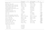
The comparison between patients with and without PH is summarized in Table 4. Statistically significant differences were observed in ACA positivity, uric acid levels, NT-proBNP levels, DLCO, ECG axis deviation, TRV, and RA area. Notably, 8 of the 11 PH cases (72.72%) were classified as WHO functional class I or II.
Comparison of clinical, functional, and laboratory characteristics between SSc patients with and without pulmonary hypertension (PH).
| PH group(n=11) | No PH group(n=74) | p | |
|---|---|---|---|
| Women, n (%) | 9 (81.81%) | 68 (91.89%) | 0.275 |
| Age (years), mean (SD) | 69.90 (± 9.70) | 67.00 (± 12.00) | 0.222 |
| Age at diagnosis (years), mean (SD) | 50.00 (± 13.70) | 51.70 (± 14.20) | 0.358 |
| Disease duration (years), mean (SD) | 19.90 (± 11.20) | 15.00 (± 8.70) | 0.109 |
| Limited cutaneous SSc, n (%) | 11 (100%) | 64 (88.90%) | 0.303 |
| Raynaud's phenomenon, n (%) | 11 (100%) | 72 (97.29%) | 0.757 |
| Telangiectasia, n (%) | 9 (81.81%) | 47 (63.51%) | 0.403 |
| Interstitial lung disease, n (%) | 0 (0%) | 15 (23.27%) | 0.101 |
| Pulmonary emphysema, n (%) | 1 (10.00%) | 0 (0%) | 0.147 |
| ACA, n (%) | 11 (100%) | 34 (45.94%) | <0.001 |
| ATA, n (%) | 0 (0%) | 11 (14.94%) | 0.196 |
| Serum uric acid (mg/dL), mean (SD) | 6.96 (± 1.72) | 5.10 (± 1.77) | <0.001 |
| Serum NT-proBNP (pg/dL), mean (SD) | 1072.27 (± 1140.28) | 546.71 (± 1015.90) | 0.002 |
| FVC %, mean (SD) | 92.20 (± 17.40) | 92.25 (± 15.28) | 0.496 |
| DLCO %, mean (SD) | 43.08 (± 15.64) | 65.64 (± 16.72) | <0.001 |
| EKG axis deviation, n (%) | 2 (18.18%) | 0 (0%) | <0.001 |
| Points from Step 1, mean (SD) | 370.09 (± 46.12) | 321.52 (± 20.08) | <0.001 |
| TRV (m/s), mean (SD) | 3.53 (± 0.85) | 2.50 (± 0.45) | 0.012 |
| RA area (cm2), mean (SD) | 21.84 (± 10.15) | 15.11 (± 3.94) | <0.001 |
| WHO functional class (FC) | |||
| FC I, n (%) | 3 (27.27%) | ||
| FC II, n (%) | 5 (45.46%) | ||
| FC III, n (%) | 2 (18.18%) | ||
| FC IV, n (%) | 1 (9.09%) | ||
In the bivariate analysis (Table 5), a higher tricuspid regurgitation velocity (TRV) and a lower DLCO were significantly associated with the presence of PH. Additionally, higher scores in both Step 1 and Step 2 of the DETECT algorithm were also significantly associated with PH. In the multivariable analysis (Table 6), TRV remained an independent predictor of PH (OR=11.57; 95% CI: 1.287–103.976; p=0.029), while DLCO was inversely associated with PH (OR=0.89; 95% CI: 0.797–0.987; p=0.028).
Bivariable analysis of the odds ratio (OR) for PH development.
| Variable | OR | IC 95% | p |
|---|---|---|---|
| Telangiectasia | 0.15 | 0.009–2.373 | 0.178 |
| Serum uric acid | 1.49 | 0.897–2.466 | 0.124 |
| Serum NT-proBNP | 1.000 | 0.999–1.001 | 0.575 |
| DLCO %, mean | 0.93 | 0.872–0.991 | 0.025 |
| Points from Step 1, mean | 1.11 | 1.029–1.192 | 0.006 |
| RA area (cm2), mean | 0.92 | 0.761–1.113 | 0.391 |
| TRV (m/s), mean | 33.28 | 2.372–466.948 | 0.009 |
| Points from Step 2, mean | 1.14 | 1.024–1.272 | 0.017 |
Multivariable analysis of the odds ratio (OR) for HP development.
| Variable | OR | IC 95% | p |
|---|---|---|---|
| Sex (woman) | 112.49 | 0.383–33,066.189 | 0.103 |
| Age (years) | 0.99 | 0.894–1.090 | 0.804 |
| DLCO % | 0.89 | 0.797–0.987 | 0.028 |
| TRV (m/s) | 11.57 | 1.287–103.976 | 0.029 |
Abbreviations: SD, standard deviation; Ref, reference threshold; NT-proBNP, N-terminal fragment of the brain natriuretic peptide; FVC, forced vital capacity; DLCO, diffusion capacity for carbon monoxide; EKG, electrocardiogram; TTE: transthoracic echocardiogram, TRV, tricuspid regurgitation velocity; RA, right atrium; RHC, right heart catheterization; PH, pulmonary hypertension; TE, thromboembolic.
In this study, we evaluated the clinical utility of the DETECT algorithm for the identification of pulmonary hypertension (PH) in a real-world cohort of systemic sclerosis (SSc) patients without a prior PH diagnosis.
The most common epidemiological profile in the cohort consisted of women in their sixth and seventh decades of life, predominantly with the limited cutaneous form of SSc and a mean disease duration of approximately 15 years. This profile is consistent with previous reports. In the study by Avouac et al.,15 PH was evaluated in two cohorts of SSc patients—one French and one Italian— where most patients were women, with mean ages of 57 and 62 years, respectively. The limited cutaneous form was significantly more frequent than the diffuse subtype, and the mean disease duration was approximately 13 years. A meta-analysis included in that publication also analyzed four additional cohorts from the United Kingdom, France, the Netherlands, and Australia,16–19 showing similar baseline characteristics.
Notably, 47% of patients in our cohort were ACA negative, despite the predominance of the limited form of SSc. This observation is consistent with findings by Höpner et al.,20 who analyzed the autoantibody profile in 372 SSc patients and their clinical associations. In that study, anti-Ro52 antibodies were significantly associated with the presence of PH. Moreover, PH was identified in 21.4% of patients with anti-Th/To antibodies, compared to 14.5% of those with ACA positivity.
An interesting finding in our cohort was the mean DLCO of 61.64%±18.57, observed despite the relatively low prevalence of confirmed PH. Corzo et al.21 described a similar subgroup of SSc patients with mildly reduced DLCO (mean 65.60±10.60%), predominantly with limited SSc and ACA positivity, but without evidence of interstitial lung disease or emphysema.
In our cohort, interstitial lung disease (ILD) was present in 17.60% of patients, while emphysema was reported in a single case, and no patient had documented thromboembolic pulmonary disease. These findings suggest that the observed DLCO reductions cannot be fully explained by parenchymal or thrombotic pulmonary damage. Regarding the serological profile, anti-topoisomerase I antibodies were identified in 11 patients (12.90%), all of whom were ACA negative. Although ATA are classically associated with the diffuse cutaneous form of SSc, our cohort was predominantly composed of patients with limited cutaneous disease, which may reflect either atypical serological-phenotypic associations or the limited sample size.
It has been proposed that a mild reduction in DLCO may reflect early pulmonary arterial vasculopathy, particularly in patients with shorter disease duration, as PH generally develops after 10–20 years of SSc evolution. Nonetheless, isolated DLCO reduction is not considered a strong prognostic marker, unlike more severe decreases (< 45–55%), which have been associated with a higher risk of PH. In this context, the DETECT study demonstrated a low prevalence of PH among patients with DLCO>60%, suggesting that the reduction observed in our cohort may reflect early microvascular involvement, although its prognostic significance remains uncertain and warrants longitudinal follow-up.
In our population, higher DLCO values were inversely associated with the presence of PH, supporting the hypothesis that preserved gas exchange capacity may serve as a predictive factor. This observation aligns with the 2022 ESC/ERC Guidelines, which emphasize DLCO reduction as a frequent finding in SSc-associated PH. Launay et al.22 incorporated DLCO into a cluster analysis of 200 patients, identifying four phenotypes with markedly different three-year survival outcomes, thereby reinforcing the prognostic role of DLCO. Similarly, Allanore et al.23 demonstrated that DLCO<70% and <60% were both significant predictors of future PH development in a longitudinal cohort of 101 patients.
In our study, elevated tricuspid regurgitation velocity (TRV) emerged as a strong predictor of PH, showing statistical significance in both bivariate and multivariate analyses (OR=11.570; 95% CI: 1.287–103.976; p=0.029). TRV is widely recognized as a key echocardiographic parameter in PH risk stratification among SSc patients. According to the 2022 ESC/ERS Guidelines,1 based on findings by D’Alto et al.,24 a TRV≥2.9m/s is independently associated with PH development, whereas a TRV<2.8m/s—when no other echocardiographic abnormalities are present—makes PH unlikely.
Following the application of the DETECT algorithm, PH was confirmed in 11 patients (12.9%) within our cohort. This prevalence is higher than that reported in some previous studies and may be partially explained by the relatively long mean disease duration observed in our population (15.7 years), which is a known risk factor for PH development in SSc.
Humbert et al.5 compared two cohorts of SSc patients undergoing PH screening: one assessed through routine clinical practice and the other enrolled a systematic screening program. In that study, TTE was performed in 570 SSc patients without prior PH diagnosis or severe pulmonary impairment, excluding secondary PH. PH was suspected in 33 patients (5.78%), and confirmed by RHC in 18 (3.16%). Notably, patients identified through systematic screening exhibited less advanced pulmonary vascular disease, as evidenced by a greater proportion in NYHA functional classes I and II, lower pulmonary arterial pressure and resistance, and higher cardiac output. Survival during follow-up was significantly better in the systematic screened cohort compared to the one assessed through routine care.
Like our findings, Hachulla et al.17 conducted a comparative analysis of SSc patients with and without PH, and reported significant differences in serum uric acid, NT-proBNP levels, ACA positivity, DLCO, TRV, and right atrium area—reinforcing the value of these biomarkers for early PH detection. However, their cohort, also showed significant differences in sex distribution, age, and the presence of telangiectasia, which were not observed in our study.
In our study, diagnostic accuracy parameters for the DETECT algorithm could not be determined, as right heart catheterization was not performed in all patients. In contrast, the original DETECT study,12 which included RHC in the entire cohort, reported a sensitivity of 96%, specificity of 48%, a positive predictive value (PPV) of 35%, and a negative predictive value (NPV) of 98%.
A post hoc analysis25 of this study, the updated PH definition from the 2022 ESC/ERS guidelines1 was applied, setting the threshold at a mean pulmonary arterial pressure (mPAP)>20mmHg instead of >25mmHg, as used when the algorithm was developed. The results of this new analysis showed lower sensitivity (88.2%) and NPV (89.7%) than in the original study but improved specificity (50.8%) and PPV (46.9%).
Several studies have assessed the diagnostic performance of the DETECT algorithm, consistently reporting high sensitivity but limited specificity. For instance, Young et al.26 applied the algorithm to a cohort of 68 SSc patients, including 10 with confirmed PH, and reported a sensitivity of 100%, specificity of 29%, PPV of 20%, and NPV of 94%.
In the study by Guillén del Castillo et al.,27 which included 83 SSc patients, the DETECT algorithm demonstrated a sensitivity of 100%, specificity of 42.9%, PPV of 68.6%, and NPV of 100%. Similarly, Hachulla et al.17 assessed three screening algorithms in a cohort of 73 SSc patients, where DETECT again achieved high sensitivity (100%), and NPV (100%). The performance of DETECT in this study was comparable to that of the Australian Scleroderma Interest Group (ASIG) algorithm and superior to the ESC/ERS criteria.
Erdogan et al.28 compared the DETECT, ASIG, and ESC/ERS screening algorithms using both PH definitions: mPAP>20mmHg and >25mmHg. Under the >20mmHg cutoff, DETECT showed a sensitivity of 80%, specificity of 82%, PPV of 59%, and NPV of 93%. When using the >25mmHg threshold, sensitivity increased to 100%, while specificity decreased to 73%, with a PPV of 26% and NPV of 100%. Overall, DETECT performed similarly to the ASIG algorithm and outperformed the ESC/ERS criteria under both definitions.
Compared to previous PH screening strategies, the DETECT algorithm facilitates earlier identification of PH in SSc patients, while maintaining a low false-negative rate. Reported sensitivities were 96% in the original study and 88.2% in a post hoc analysis, applying the updated PH definition.13,25 Its two-step structure and reliance on routinely available clinical variables enhance its practicality for implementation in everyday clinical settings.
Despite its high sensitivity, the DETECT algorithm has a limited specificity, with a false-positive rate of 65% reported in the original study,23 highlighting the potential need for refinement. The incorporation of additional biomarkers—such as endothelin-1, vascular endothelial growth factor (VEGF), and adhesion molecules (VCAM-1, ICAM-1)—may improve its diagnostic performance, given their established associations with SSc-related PH.29
Although the DETECT algorithm shows limited specificity across multiple studies, its overall performance remains comparable to—even superior to—other existing PH screening strategies. Its principal strength lies in its high sensitivity, which minimizes the risk of missed diagnosis and facilitates early therapeutic intervention.
Our findings suggest that the DETECT algorithm may also be applicable to patients with DLCO>60%, thereby extending its utility beyond the initially proposed threshold. Although this cutoff was initially intended to identify a higher risk population, its exclusionary role in patients with DLCO>60% remains under debate and warrants further evaluation.
In our cohort, where the mean DLCO was 61.64±18.57%, PH was identified in a notable proportion of patients. This finding supports the potential value of implementing the DETECT algorithm even in individuals with relatively preserved DLCO. However, further investigation is warranted, as the prevalence of PH in this subgroup remains lower than in the high-risk population for which the algorithm was originally designed.
Standardizing the DETECT algorithm in clinical practice may aid in early identification of PH, particularly in patients with mild symptoms, by optimizing the indication for complementary diagnostic tests. From a clinical standpoint, its implementation offers a structured and reproducible approach to PH screening in SSc. Future research should focus on validating its reproducibility across diverse populations and evaluating its long-term impact on prognosis and cost-effectiveness.
This study has several limitations. Being a single-center analysis, the generalizability of our findings may be limited. The exclusion of patients with advanced chronic kidney disease also restricts the extrapolation of results in this potentially high-risk subgroup. Moreover, the sample size may have limited the statistical power to detect significant associations in the multivariable model. Importantly, since RHC was not performed in all patients, diagnostic accuracy metrics could not be calculated. Lastly, the cross-sectional design prevents assessment of causality or longitudinal outcomes related to early PH detection.
ConclusionThe implementation of the DETECT algorithm in clinical practice represents a valuable approach for PH screening in SSc patients. Our findings support a meaningful correlation between the algorithm's variables and PH diagnoses confirmed by RHC. Beyond its diagnostic value, the algorithm holds potential clinical relevance by enabling earlier detection and possibly improving long-term outcomes.
Further studies are warranted to validate its applicability across diverse populations and, most importantly, to evaluate its impact on the long-term prognosis of SSc patients.
FundingGGA is a predoctoral researcher at the Rio Hortega Program of the ISCIII, Reference #CM24/00240.
Conflict of interestThe authors declare that they have no conflict of interest.
The authors would like to thank Dr. Iván Castellví for his time, guidance, and generosity during the preparation of this work, as part of the Mentoring Program of the Spanish Society of Rheumatology.


