


The primary vasculitidies are complex diseases with varied clinical manifestations, which may be common to those present in multiple diseases. The antineutrophil cytoplasm autoantibodies (ANCA) led to a revolution in the diagnosis and research of these diseases, being the first and so far, the only biomarkers for 3 of these diseases, which affect small caliber vessels. From their description, much progress has been made, but there are still gray or misunderstood areas regarding their best use in the clinic. This can lead to errors as making a positive test synonym for vasculitis, or to overestimation of its importance. This review will address aspects such as nomenclature, employment in the diagnosis and monitoring of vasculitis, their presence in other diseases, their methods of detection, and finally, some comments on other potential biomarkers in vasculitis.
Las vasculitis primarias son patologías complejas, con manifestaciones clínicas variadas y proteas, las que pueden ser comunes a las que se presentan en múltiples enfermedades. Los anticuerpos anticitoplasma de neutrófilo (ANCA) constituyeron una revolución en el diagnóstico y la investigación de estas enfermedades, al ser el primer, y hasta ahora único, biomarcador para 3 de estas patologías que afectan vasos de calibre pequeño. Desde su descripción, mucho se ha avanzado, pero aún hay aspectos poco claros o mal entendidos en cuanto a su mejor uso en la clínica. Ello puede llevar a errores como hacer de esta prueba un sinónimo de la existencia de vasculitis, o a sobreestimar su relevancia. Esta revisión abordará aspectos como su nomenclatura, el empleo en el diagnóstico y seguimiento de las vasculitis, su presencia en otras enfermedades, algunos comentarios respecto de sus métodos de detección, y finalmente, observaciones respecto de otros potenciales biomarcadores en las vasculitis.
“The fact that something does not work as expected does not mean it is not useful”. Thomas Alva Edison.
After more than 25 years there is no doubt on the usefulness of antineutrophil cytoplasm antibodies (ANCA). However, as in any human activity, their use, including indications, performance, and interpretation, is what determines their real value. They are of such importance that, to date, constitute the only biomarkers in a heterogeneous group of diseases of difficult diagnosis and complexity regarding treatment and monitoring of vasculitidies. More than 150 years since the first recognition of these diseases,1 and the time elapsed between the firm description of ANCA as a diagnostic marker for cases of Wegener's granulomatosis and Polyangiitis,2 which are still considered rare entities, there is no other test at the moment that helps us in the same magnitude. Like any laboratory study, it is subject to many variables, among which is the detection methodology. In this manuscript I will strive to elaborate on the issues reported in the summary.
Antineutrophil Cytoplasm Antibodies in DiagnosisIt is an incontrovertible truth: ANCA are useful in diagnosing certain primary vasculitidies; in fact, it is the most commonly performed test except in certain diseases, such as giant cell arteritis. Since the original paper by van der Woude and Wiik, it was clear that in a rare condition such as GPA, ANCA detected by indirect immunofluorescence were highly specific for this disease.2 That publication identified the association of perinuclear pattern (P-ANCA) with pauciimmune nephritis3,4 and the identification of relevant antigens for each pattern of immunofluoresence,5,6 and the development of solid phase methods for the detection of such antigens, combined with the former, established its utility as published by the joint European study group in 1998.7 One of the highlights was to establish that 3-GPA pathology, microscopic polyangiitis (MPA), and vasculitis limited to the kidneys (in the presence or absence of rapidly progressive glomerulonephritis)–are strongly associated with these antibodies and that the performance of both tests increases significantly its sensitivity and specificity, compared with control patients in the differential diagnosis may show clinical features similar to those observed in these pathologies. With that, some indications were established by a group of experts, which have served as a general guideline to the application of these antibodies (Table 1),8 although in this recommendation, it is stated that inmunofluoresence (IFI) is a scrutiny test, which may be done after performing ELISA, in case the latter is positive. This despite the fact that the diseases mentioned above, which has a more positive pretest possibility, demonstrated that the simultaneous performance of both methods provides a better performance. This leads to a fundamental question: who should request ANCA? Two studies published a decade ago showed that indiscriminate application carries a high percentage of false positive, low positive, and negative predictive values, which was directly related to medical specialties. The first study showed that even some experts who requested ANCA had poor yield.9 Subsequently, another publication, which considered only those results confirmed by ELISA as positive ANCA, and performed a screening of its application according to this point, demonstrated that it was possible to decrease false positives and especially to increase predictive negative values.10 This poor performance of ANCA, even in specialized centers, has been observed in our institution as well. Results published so far in abstract form with data from the National Institute of Medical Sciences and Nutrition in México11 for a period of 12 months, analyzed the performance of each method and compared the results against defined diagnoses (Table 2), particularly groups of primary ANCA associated vasculitis, and another performed through immunofluresence (only 3% of cases within this group also included determination through ELISA for antigenic specificities), encompassing gastrointestinal and hepatic disease that has been associated with positivity in IFI. In connection with the group of vasculitis, the dispersion is clearly seen, similar to the studies presented above, with lower positive predictive values but similarly negative. Even taking into account the specific test using direct ELISA, the results are moderate, suggesting that in a tertiary care center, it is convenient to redefine the use of these tests in their current form. From this data, it has been suggested that the establishment of screening “policies” subject to certain indications is favorable when deciding to request ANCA. Recent studies suggest that this is possible without sacrificing accuracy of the evidence regarding the potential loss of patients with diseases clearly associated with these antibodies. A New Zealand tertiary center evaluated 1127 tests, of which only one third were guided by the criteria proposed in 1999. Of that percentage, all tests were positive in all cases except one who had infectious endocarditis, the diagnosis was associated with ANCA vasculitis. The rest of the entries, including ocular or orbital disease, liver disease or inflammatory bowel disease, eosinophilic granulomatosis with polyangiitis (Churg–Strauss)– not in the proposed indication – had very few positive cases, concluding that in such situations ANCA are useless as a diagnostic test. Moreover, following the directions for application proposed means that, exceptionally, the diagnosis of primary vasculitis is certain.12 Prospectively, the Oxford group showed that the application of the guidelines increased the yield of positive tests, while rejected those tests that did not meet this control and no cases of primary vasculitis associated with ANCA were detected in the 8 month follow-up.13
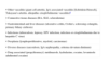
Clinical Indications for Antineutrophil Antibody Performance.
| Glomerulonephritis, especially rapidly progressive |
| Lung hemorrhage, especially lung–kidney syndromes |
| Skin vasculitis with systemic affection |
| Multiple lung nodules |
| Chronic destructive upper airway disease |
| Sinusitis or chornic otitis |
| Tracheal stenosis, subglottic stenosis |
| Mononeuritis multiplex or peripheral neuropathy |
| Retroorbital masses |
Properties of the ANCA Test (IFI, ELISA, and Together) in Patients With an ANCA Associated Vasculitis as Requested by Any Department of a Third Level Hospital in Mexico City.
| Method | Sensitivity, % | Specificity, % | PPV, % | NPV, % |
| IFIa | 86 | 18 | 13 | 86 |
| ELISAb | 54 | 82 | 64 | 69 |
| IFI+ELISAc | 86 | 23 | 14 | 92 |
IFI, indirect immunofluorescence.
But are there other ANCA positive diseases? Is the term ANCA-associated correct? Table 3 cites other diseases that have reported as ANCA positive, noting particularly those where their presence is more common, their association with clinical manifestations similar to those seen in ANCA positive vasculitis or those that are of particular interest in certain situations, particularly rheumatic diseases, where there is a high prevalence of infectious disease.14 In connection with inflammatory bowel disease, most of the information is retrospective, but the reactivity found is directed to many antigens and not against the neutrophil myeloperoxidase (MPO) or proteinase 3 (PR-3). In any case, in general, when ANCA is positive in these diseases, it is more common in ulcerative colitis than in Crohn's disease, where its activity is questionable under these conditions.15 Interestingly, in Crohn's disease with colonic involvement, ANCA are positive more frequently than in those without such an affection.16 As for their relationship with drugs or other substances, particularly cocaine, ANCA seem to recognize the antigen and some cases may exist that also have other autoantibodies in their serum. In fact, the ANCA reaction may be directed against several enzymes in neutrophils in conjunction with anti-histone antibodies, anti-beta 2 glycoprotein 1 and/or anticardiolipin antibodies, identified in these patients.17,18 I will refer to a recently identified situation, particularly in view of the likely increase of this problem in times to come and in particular social contexts. In 2006, although reported until 2009, several patients were identified with cutaneous vasculitis in peculiar locations and changes in mucous membranes, in some cases with severe neutropenia, which were ANCA positive, including reactivity against PR-3, MPO, and many other neutrophil antigens (elastase, cathepsin G, lactoferrin), and several of them also against cardiolipin, lupus anticoagulant, low complement levels or marginally cryoglobulins which has been the common denominator of cocaine adulterated with levamisole. The latter causes agranulocytosis, arthralgias, hemolytic anemia, hepatosplenomegaly, and characteristically, vasculitic lesions with a particular distribution in ears and cheeks, and when affecting the limbs, with large and deep ulcers. Histologically, the most frequently observed alteration is thrombotic microangiopathy, but in some cases leukocytoclastic vasculitis has been documented. The suspension of the use of cocaine and supportive measures in less severe cases is usually sufficient to obtain relief, but in serious cases steroids have been employed.19–23 It is, of course, essential in these cases to rule out infection, but a full and appropriate diagnostic approach, and the characteristics mentioned must lead to an exhaustive search of a history of cocaine use, which, as noted, is highly feasible to be found adulterated with levamisole. We have seen 2 cases (manuscript in preparation), of which one of them, despite 2 previous hospitalizations, sadly passed unsuspected. Thus, in patients with these characteristics, we must assume this problem. Other conditions, in which ANCA were recently reported positive, together with those in Table 3, are neutropenia and cyclic erythema elevatum diutinum.24,25 In the first, although the immunofluorescence pattern is cytoplasmic, the reactivity seems to be against a 60kDa antigen which is awaiting precise characterization, and in the second case against the IgA isotype which has been described in some patients with this condition. This leads to the following points: neutrophils (and monocytes as well) are full of proteins and enzymes in which, schematically, faulty regulatory mechanisms in conjunction with aberrant immune phenomena may trigger an autoimmune response, which in an appropriate environment, can become an autoimmune disease. We now know that the link of antibodies against PR-3 and MPO occurs with specific diseases and know that at some time, the MPO-ANCA have an important pathogenic potential. Nevertheless, ANCA have permeated into the diagnostic routine and the requests to the laboratory for autoimmune pathologies. As such, when clinicians of different specialties face a positive ANCA result, particularly in imprecise or vague cases, it may become even risky, due to the diagnostic implications and therefore treatment that they entail. As a result, in recent years, several authors, but particularly Wiik, have pointed to positive IFI reactivity which recognizes antigens other than MPO or PR-3, such as neutrophil specific antibodies (NSA).26 Personally, such reactivity is shared between neutrophils and monocytes, so I here propose the preferred term neutrophil-recognizing antibodies (NERA).27 In any case, regardless of the term, the aim is to avoid misinterpretation of the term ANCA, which should be reserved for those antibodies recognizing MPO and PR-3 and they have, as demonstrated, specific associations, particularly with GPA, MPA, kidney limited vasculitis and to 30%–50% of patients with Churg–Strauss syndrome (CSS).28,29 This proposal would include the association of these antibodies that recognize antigens other than PR-3 and MPO, but give an immunofluorescent pattern having neutrophils as substrate, with pathologies of the liver, intestine, or those related to the use of drugs. In 2008, Kain et al. described a high frequency of antibodies against protein-2 of the lysosomal membrane (LAMP-2), which since 1995 had been described as one of the antigens recognized by ANCA. In another article, by extending their remarks, they described the presence of antibodies against LAMP-2 in 78 of 84 patients with necrotizing Pauci-immune focal segmental glomerulonephritis with a cytoplasmic IIF reactivity pattern, and the other 6 patients, in which there were no antibodies against LAMP-2, corresponded to patients in remission.30 This figure was higher than that in those patients found positive for antibodies to PR-3 and MPO. Additionally, the antibodies generated in rabbits after immunization with human LAMP-2 were able to produce, in Wistar-Kyoto rats, glomerular lesions similar to those seen in patients. These antibodies were capable of activating neutrophils and endothelial cells in vitro by promoting apoptosis, and also cross reacted to an epitope present in an adhesin-negative fimbriae (FimH). By immunizing mice with recombinant FimH they showed that these antibodies also recognized human LAMP-2, without recognizing MPO or PR-3. Finally, retrospectively, using information obtained from primary care physicians of 9 of their most recent 13 patients with renal disease and establishing the presence of gram-negative bacterial disease 12 weeks prior to the establishment of the renal disease. With these data it is suggested that as seen in renal pathology, ANCA directed against LAMP-2 are more prevalent and specific than those against MPO and/or PR-3, having a potential pathogenic role. Their experiments also support the hypothesis of crossreactivity between infections and the generation of these diseases. Of course, this was very striking in view of the lack of reactivity against this antigen in original descriptions of the search for antigens responsible for neutrophil reactivity of nearly 30 years. These observations have not been confirmed in 2 groups of patients in the U.S. By expanding the search for anti-LAMP-2 in cohorts of the Massachusetts General Hospital and the University of North Carolina at Chapel Hill, Roth et al.31 found reactivity against LAMP-2 in 21% of their groups ANCA, which was not specific, since 16% of patients with urinary tract infections also had antibodies against this antigen. Additionally, levels of antibodies to PR-3 and MPO in these sera were much higher than those found against LAMP-2. Thus, the controversy in the case of ANCA-associated vasculitis remains, and at this time it is not advisable to search for antibodies against this antigen routinely that in addition to affordability is not available and may not offer significant advantages in the clinical context. In contrast, there is a recent report of the presence of anti-LAMP-2 in IgA-associated vasculitis (Schönlein-Henoch)32 that warrants further studies in order to be confirmed as a biomarker in this vasculitis.
Other Diseases Described With Positive ANCA.
| • Other vasculitis (giant cell arteritis, IgA associated vasculitis [Schönlein-Henoch], Takayasu's arteritis, idiopathic crioglobulinemic vasculitis)a |
| • Connective tissue diseases (RA, SLE, scleroderma) |
| • Gastrointestinal and liver diseases (ulcerative colitis, Crohn's, sclerosing colangitis, primary biliary cirrhosis) |
| • Infections (tuberculosis, leprosy, HIV infection, infection or crioglobulinemia due to hepatitis C virus) |
| • Neoplasia (lymphoproliferative, myeloid, carcinomas) |
| • Diverse diseases (sarcoidosis, IgA nephropathy, eritema elevatum diutinum) |
| • Drug associated (propyltiouracyl, metilmazole, hydralazine, cocaine, levamisole adulterated cocaine) |
One of the most desirable properties of a serological test is its performance in monitoring patients. A substantial accumulation of information has emerged since the late eighties, when the Groningen group found evidence that, in particular ANCA using capture ELISA, could be a reliable screening method to predict relapse and guide treatment decisions.33,34 This could not be replicated by other groups or even used to evaluate other immunoglobulin isotypes other than IgG or its subclasses.35,36 A review showed years ago that the use of ANCA with this intention was unacceptable.37 This was reinforced by a well-known prospective cohort study of etanercept in GPA, which, with data from 156 patients, determined the time of referral related to the decrease in ANCA as measured by ELISA 2 against mature and promolecuea PR-3, and the relapses that followed sustained remissions as well as the relationship of these with increased levels of ANCA.38 Definitions of increases and decreases in ANCA were arbitrary, as has happened in all studies. Of those patients positive for PR3-ANCA who were managed and subsequently remmited, there was an increase in one third of patients with either of the 2 methods. However, this increase was not followed by clinical relapses, and in some cases there were patients in whom the onset of clinical manifestations occurred 1 year after the increase in the levels of PR3-ANCA. There were also patients in whom there was no increase of PR3-ANCA before clinical relapses. Moreover, in patients negative at enrollment and who remained with elevated ANCA, half had a relapse of ANCA elevation prior to any of the 2 methods used for detection. A meta-analysis concluded that the most recent value of serial measurements of ANCA in patients in remission is equally limited. Neither the increase nor its persistence in subjects who achieved remission is highly predictive of relapses.39 I have mentioned IFI and ELISA several times. Is there a single ELISA? Does this affect the performance of ANCA in both diagnosis and follow-up? Space does not permit further discussion in the matter, but it is possible to comment that there are discrepancies between different ELISA methods, either direct or capture, used to detect antibodies against MPO or PR-3, which can be influenced by the chosen cut points.40 A cut point above 2 standard deviations using healthy subjects as controls gives a high specificity, but even 20% of patients with diseases that can potentially be confused with ANCA-associated vasculitis may be positive. If this cutoff is raised, some patients, especially those with less florid manifestations, such as a limited GPA, and constitutes something which is essential to conduct further studies to look for other causes of their symptoms or seek histological damage may lose positivity in the ELISA, and the possibility that the outcome is positive having other pathology in the differential diagnosis is reduced from 20% to 5%. In addition, it provides compared to healthy subjects, almost absolute27 specificity. Thus, knowing some details of the studies and tests commercially offered locally is of great importance. Comparisons of different types of ELISA give similar results, although, initially, in monitoring its use, through both direct and capture methods, the latter being more sensitive appeared to be superior in predicting a relapse. Other developments in enzyme immunoassays have been tested, but in general, almost all the tests have good specificity and variable sensitivity. A recent development concerns the location of the molecules (whose composition was not described in detail in the publication) of the antigen targets, which appear to increase the sensitivity of the solid assay phase, keeping relevant epitopes on antigens and fixed for specific41 recognition by antibodies. Studies with platforms such as Luminex are, of course, very attractive, but at the moment extremely expensive for routine implementation and therefore not recommended for use.42 There are several causes of variability in the use of ANCA in monitoring patients, including the definition of increased levels, the frequency measurements must be made to meet the goal of prediction, the clinical status definitions (although this is increasingly solved with the use of measures such as BVAS), the method used in each center, with its inherent characteristics, which would be highly desirable by clinicians, and, as we have seen, the characteristics of the antigen coupled to the detection method employed.43 Overall there is evidence to support the conclusion that in PR3-ANCA positive patients, it is safer to make the change of remission induction therapy to the maintenance phase when antibodies are negative. Disease-free survival was superior to those PR3-ANCA positive patients when switched from cyclophosphamide to azathioprine.44 In this and subsequent studies, one of them with information from the cohorts the European vasculitis group therapeutic studies, PR3-ANCA positive cases are more likely to relapse than MPO-ANCA positive, and therefore, it is desirable to closely monitor the former.45 Another situation is, of course, to identify the serological behavior of each patient. Thus, some patients present minor variations of ANCA levels, but others, at least at the time of clinical manifestations of activity, have elevated levels of ANCA and their behavior is somewhat constant or stereotyped between clinical and serology data. Accurate monitoring can help determine if a patient tends to raise her/his levels just before or during relapses, and whether this has influence on clinical behavior. Thus, the latter remains the main determinant of changes in treatment decisions, even despite the fact that C-reactive protein (CRP), as a recently described highly sensitive development marker of inflammation, and which is certainly not unique in these conditions, can be raised concurrently in the presence of relapses.46 At present, even in the situations mentioned above, the clinical findings remain the major determinants of therapeutic changes in these conditions.
Antineutrophil Cytoplasm Antibodies in Clinically Defined Situations: An ExampleAt least 10% and even up to 30% of patients with positive ANCA develop kidney failure which warrants replacement of renal function. It is known that the outcome of transplant recipients and their survival are equal to that of other diseases.47 Despite immunosuppressive treatment regimens, up to 20% of transplanted patients relapse, which occurs on average after 2 to 3 years. One of the frequently asked questions is related to the association between ANCA levels and the time of transplantation. Despite the widespread notions, it is known that once clinical remission is achieved, transplantation is successful, regardless of ANCA48 levels. Since then, according to the above, it is better if the procedure is performed with negative antibody levels, but positivity does not necessarily imply a worse prognosis in terms of renal survival. In any case, relapse in transplanted patients has been observed when the serum is positive for ANCA. Furthermore, extrarenal relapses occur in these patients in up to half of the cases and are not related to the time since grafting. Is there a difference between MPA and GPA in terms of these characteristics in renal transplant recipients? Recently, it has been reported that renal transplantation is lower in patients with MPA than GPA, although this is not significant,49 but this situation merits monitoring, particularly at a time when transplant immunosuppressive regimens have changed and favor the use MMF and tacrolimus. In summary, although it seems desirable to transplant patients when they are negative for ANCA, their presence should not determine the time of transplantation, with clinical activity being the main determinant, and maintaining appropriate monitoring for possible relapses, despite regimes of immunosuppression to prevent rejection that are appropriate and drug levels are optimal in this patient group.
Other Markers in VasculitisI commented on the usefulness of ANCA and over the years it has become clear that they are an important diagnostic biomarker in small caliber vasculitis, particularly in 2 (GPA and MPA), and also helped to clarify that these 2 diseases are responsible for most of the syndromatic entities: rapidly progressive glomerulonephritis syndrome and diffuse alveolar hemorrhage. Furthermore, although there is reasonable evidence of their pathogenic role, the initial expectations of ANCA as an unequivocal biological marker to predict relapse and guide therapeutic decisions to be specific enough to explain manifestations of the disease, compared with that seen in other diseases or complications of treatment, including infections, remain unfulfilled. In this final part I want to comment briefly on progress in the search for other biomarkers that should complement or surpass ANCA in these areas. These conditions have many actors, either as triggers, as victims or both. To the extent that questions are answered more arise in the search for relevant molecules in these conditions, both needed to be sensitive and specific, and consequently useful. Table 4 lists several that have been described over the last decade. One target is the endothelium. While circulating endothelial cells increase in activity phases of the diseases associated with ANCA, it appears to be clearly established to damage,50 while endothelial microparticles, those derived from early lesions, appear to increase rapidly in some cases even before clinical manifestations, and return to normal when the disease is in remission.51 The participation of B lymphocytes in the pathogenesis has naturally led to the search for the related markers. It has recently been reported that levels of B cell activating factor (BAFF) in serum were higher in GPA patients than in healthy controls, but it was negatively correlated with ANCA levels and even levels of BAFF were higher in patients with negative ANCA in addition to the fact that there was neither a correlation with other measures of activity such as clinical indices or CRP, nor increased levels at the time of recovery.52 Another group studied this, where patient levels of BAFF were high compared with those with MPA or Churg–Strauss. In the same study, markers of endothelial activation and VCAM-1 also increased during relapses, but in terms of BAFF levels there was no greater difference between the different stages of the disease.53 The latter is somewhat surprising, but it is possible that it constitutes a contraregulating mechanism to greater lymphocyte activation. As for markers of T cell activation, the levels of IL-17 and IL-23 have also been found increased, while those of the latter were related to activity and ANCA.54 Interestingly, the fall in the levels of both was not significant after therapy, so the persistent levels of these IL could identify a subgroup with a greater tendency to relapse. One of the most important aspects is the distinction between clinical signs of disease or infection. Recently, a Japanese group reported that the measurement of soluble receptor activator in myeloid cells 1 (TREM-1) in patients with MPO-ANCA positive vasculitis, in remission and with inactive infections, could be useful to distinguish between infection and activity. The relationship between TREM-1/creatinine was higher in patients in remission with infections, similar to that seen in patients with pyelonephritis and different from other groups, including one with chronic renal failure.55 Finally, other autoantibodies in these diseases may be important to explain certain pathogenic phenomena with clinical counterparts. The presence of antibodies against plasminogen studied in British and Dutch cohorts, and present both in 25% of patients, explains at least partially thrombotic complications in these patients. In those with such antibodies, their renal biopsies showed more fibrinoid necrosis and cellular crescents in glomeruli, which correlated with lower renal56 function. In Churg–Strauss, on the other hand, eotaxin 3 levels have been elevated during disease activity, and it also apparently occurs only in this condition and not in other hypereosynophillic syndromes; it seems a specific, sensitive, and promising marker in this disease.57 The development of new technologies such as protein microarray ELISA or Luminex technology could in a short time, identify useful biomarkers in these entities. The first has been employed in the sera of patients from the RAVE study cohort and in the near future will present results (Monach PA, personal communication). Meanwhile, this same author and the RAVE study group have reported that vascular inflammatory markers such as E-selectin, did not correlate with levels of activity, whereas other tissue regeneration proteins, such as matrix metalloproteinase 3 (MMP-3), appear to be biomarkers that may better distinguish activity, although the correlation with CRP and ESR was weak.58 Angiopoietin-2, although initially thought to be a promising molecule, did not demonstrate its usefulness for discriminating remission59 or activity60 in the same group of patients. A weakness of both cohort studies from RAVE was the interval between assessments, which was 6 months, with all the variable inherent practices that clinical practice implies and further confirmation is required as well as studies in other cohorts and a precise definition between the different subpopulations of patients, and eventually their feasibility for application in routine clinical settings.
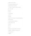
Other Potential Biomarkers in Vasculitis (Particularly Small Caliber Vessels).50–60
| • High sensitivity CRP |
| • Circulating endothelial cells |
| • Endothelial microparticles |
| • VCAM-1, ICAM-1, E and P-selectins |
| • IL-2, IL-2sR |
| • IL-18 |
| • TNF-α |
| • Osteopontin |
| • Grelin |
| • Leptin |
| • Lipocalin associates to neutrophil gelatinase |
| • Procalcitonin |
| • Antiendothelium antibodies |
| • Anti-oxidized LDL |
| • BAFF |
| • IL-23 |
| • IL-17 |
| • Eotaxin-3 (Churg–Strauss syndrome) |
| • TREM-1 (TREM-1/creatinine rate) |
| • Angiopoietin 2 |
| • MMP-3 |
In conclusion, there are several methods to detect ANCA, which although not standardized (and hardly will be in the near future), used in combination, regardless of whether it is direct ELISA, capture ELISA or newer technologies, but with a targeted clinical indication and with specificity for PR-3 or MPO, are of great diagnostic value in small-vessel vasculitis: GPA, MPA (and its kidneys limited variant), and CSC. In the latter, although present in 30%–50% of cases, they seem to define different clinical subtypes of prognosis. I should point out that, as clinicopathologic entities, and although ANCA are excellent markers to support and/or establish the diagnosis with a high pretest probability, it is desirable to have histological confirmation, particularly in difficult cases.61 There are various conditions where those antibodies hitherto called ANCA, but directed against other antigens, are also present, including newly described pathologies. It is likely that these antibodies will be named differently and for this we have proposed other terms, reserving ANCA to those seen in primary vasculitis. This is, in my view, important even when performing IFI, in an analogy with what happens with antinuclear antibodies, for detection of these antibodies can extend the diagnostic possibilities in light of the above. At present, ANCA are not related to the presence or reliably predict relapse, and there are specific clinical situations where their usefulness is yet unknown. In any case, the clinician is required to know the peculiarities of ANCA and communication with the laboratory is essential, and the latter is obliged to set cut points and revise the methodology used for detection periodically. Finally, progress in the description of new biomarkers in vasculitis is associated to ANCA and the differential diagnoses that fit in each case, unquestionable refining and defining the usefulness and limits of ANCA as a test used properly by rheumatologists.
Ethical disclosuresProtection of human and animal subjects. The authors declare that no experiments were performed on humans or animals for this investigation.
Confidentiality of Data. The authors declare that they have followed the protocols of their work centre on the publication of patient data and that all the patients included in the study have received sufficient information and have given their informed consent in writing to participate in that study.
Right to privacy and informed consent. The authors declare that no patient data appears in this article.
Conflict of InterestThe author has no conflict of interest to declare.
Please cite this article as: Flores-Suárez LF. Utilidad de los anticuerpos anticitoplasma de neutrófilo en reumatología. Reumatol Clin. 2012;8:351–7.


