


Catastrophic antiphospholipid syndrome is an infrequent disease in children, but of major relevance because of its high morbidity and mortality. We report the case of a child with digital ischaemia in whom, after aetiological screening, the diagnosis of catastrophic antiphospholipid syndrome was made.
El síndrome antifosfolípido catastrófico es una entidad infrecuente en Pediatría, pero con importante relevancia dada la elevada morbimortalidad. Se expone el caso de un niño con isquemia digital en el que, tras realizar despistaje etiológico de diferentes entidades infecciosas e inflamatorias, se llegó al diagnóstico de síndrome antifosfolípido catastrófico primario.
Catastrophic antiphospholipid syndrome (CAPS), an infrequent disease but with a mortality close to 50%,1–3 is characterised by the presence of thrombotic microangiopathy (TMA) and positive testing for antiphospholipid antibodies.4
Diagnostic criteria are contained in Table 1.5 CAPS is considered secondary when there is an underlying disease, with systemic lupus erythematosus being the most common.2,3
Diagnostic criteria of catastrophic antiphospholipid syndrome.
| Diagnostic criteria |
| *Evidence of involvement of 3 or more organs, systems or tissues |
| *Development of clinical manifestations simultaneously or throughout week 1 |
| *Lab confirmation of positivity of antiphospholipid Ac (lupus anticoagulant and/or anti-cardiolipin and/or antiβ2 glycoproteina1 to level above 40 U/L) on two or more occasions separated from one another by at least 6 weeks. |
| *Exclusion from other causes |
| Definitive catastrophic antiphospholipid syndrome |
| *Meets with the 4 criteria |
| Probable catastrophic antiphospholipid syndrome |
| *Meets with the 4 criteria, but only involvement of 2 organs, systems or tissues |
| *Meets with 4 the criteria, but with no possibility of 6-week separate confirmation from the laboratory due to death or lack of determination of onset. |
| *Presence of criteria 1, 2 and 4 |
| *Presence of criteria 1, 3 and 4, y development of a third thromboembolic event despite anticoagulation for over one week and less than one month after the second event. |
Treatment of primary paediatric CAPS is not standardized, with anticoagulation and systemic corticotherapy being recommended, together with individualised use of other therapies.1,4
Clinical observationAn 11-year-old boy with pallor, coldness and pain in his fingers and toes, of one week onset. Vasculitic palmoplantar lesions and episodes of cyanosis in fingers, coinciding with stress and exposure to cold. Abdominal pain over the last 3 months and holocraneal headache of 2 years onset, with normal cerebral MR the previous year. His mother suffered from antisynthetase syndrome.
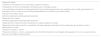
Examination revealed coldness and cyanosis in the finger pads, more intensively in the 5th finger of the right hand, 5th toe of the right foot and 3rd toe of the left foot, with no ulceration or necrosis (Fig. 1). Blood pressure, 150/115 mmHg (>p99; 4.42SD/>p99; 4.72SD), with no differences between limbs.
 Digital ischaemia and vasculitic lesions on palms of hands at onset. C and D) Improvement after one month of treatment.")
Thrombocytopenia of 109.000/mm3, with the other haematological series normal. Creatinine 0.93 mg/dL. Proteinuria 2300 mg/g, with no other alterations in urine. ANA (1/160, speckled pattern.), Ac anti-cardiolipin IgG (418U/mL), antib2GP1 IgG (104U/mL) and lupus anti-coagulant positive. Normal chest and abdominal ultrasound and angio-CT. Cortical Ischaemic lesions compatible with TMA in brain MR.
In capilaroscopy, micro-haemorrhages with changes of the capillary thickness; architecture and retained vascular density. Pale pupils with raised edge and filiform vascular network in the ocular fundus.
Epidermonecrosis in skin biopsy of the 5th toe of right foot, with no inflammatory changes or vasculopathy. Renal biopsy with mesangiolysis and double focal contours, compatible with TMA, with negative immunofluorescence.
Started treatment with aspirin, nefedipine and sidenafil, requiring an injection of iloprost one week and topical nitroglycerin due to persistence of symptoms. Following multi-organ involvement and after showing renal involvement in the biopsy made on the 6th day after hospitalization, the patient was administered with methylprednisolone bolus dose (3 days) and maintenance to 2 mg/kg/day, with progressive decline. After histological result, he began with intravenous immunoglobulin (2 g/kg) and low molecular weight heparin, progressively improving perfusion and episodes of vasoconstriction (Fig. 1), with headaches and abdominal pain disappearing and analytic tests normalizing. Due to persistent HBP, losartan was added. One month after discharge a change to oral anti-coagulation was made with acenocumarol.
After 12 months the corticoids and intravenous immunoglobulin were withdrawn, with patient being currently asymptomatic (Fig. 1) in treatment with aspirin, acenocumarol, nifedipine and losartan.
DiscussionDigital ischaemia of CAPS presents as a persistent symptom, worsening and improving for intermittent periods, when associated Raynaud’s phenomenon may occur. Pharmacological control may require combinations of vasodilator such as nefedipine, sidenafil, iloprost or bosentan. The combination of the first three, together with anti-coagulation and treatment with corticoids and intravenous immunoglobulin is effective, with the use of bosentan not being recommended due to its reported lower efficaciousness in Reynaud’s phenomenon.6 CAPS may be secondary to SLE and this is why, although our patient did not meet with the clinical or analytic criteria for its diagnosis, a high level of suspicion should be maintained because of the potential risk of developing it throughout the course of the disease.
No data exist in the literature regarding the duration of systemic treatment of CAPS, or of second line treatment in the absence of response or when there are relapses. In refractory cases in adults, good response to rituximab and eculizumab as second and third line treatments have been reported. Results appear promising1,4 despite a lack of data for children.
ConclusionPaediatric CAPS presents with multi-systemic involvement and heterogeneous clinical symptoms. A high level of suspicion is essential for carrying out early diagnosis to improve prognosis.
Conflict of interestsThe authors have no conflict of interests to declare.
Please cite this article as: Martín Pedraz L, Galindo Zavala R, Nieto Vega F, Sánchez Bazán I, Núñez Cuadros E. Isquemia digital como forma de presentación de síndrome antifosfolípido catastrófico pediátrico. Reumatol Clin. 2022;18:56–58.
This article was presented in the 7th Forum of the Spanish Society of Paediatric Rheumatology (SERPE for its initials in Spanish). Malaga, November 2018.


