


The course of sacroiliitis is characterized by inflammatory pain in the lumbosacral and gluteal region. When the condition is bilateral, it is most commonly caused by spondyloarthritis. Sometimes only 1 side is affected and, in this case, it is necessary to rule out other less common causes, such as infections or tumors, among others.
We report the case of a 49-year-old man who presented with a 1-year history of pain in right buttock. He had previously been examined in orthopedics and rehabilitation, and brought with him a technetium bone scintigraphy that showed diffuse hyperactivity in right sacroiliac joint compatible with sacroiliitis.
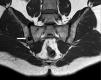
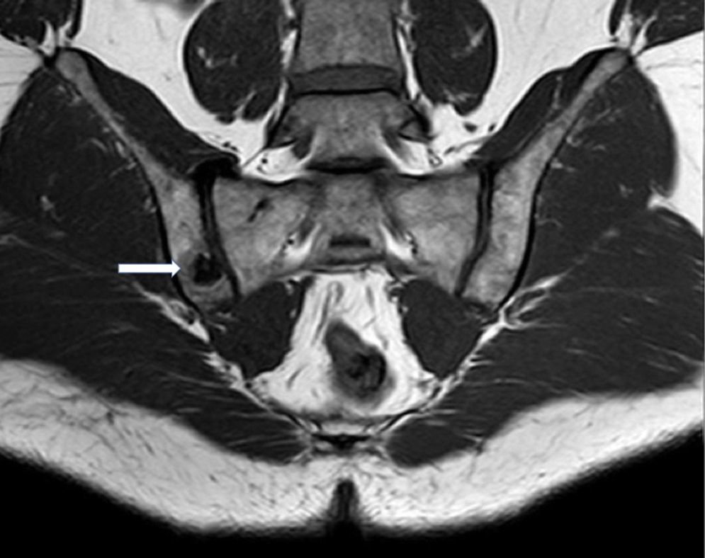
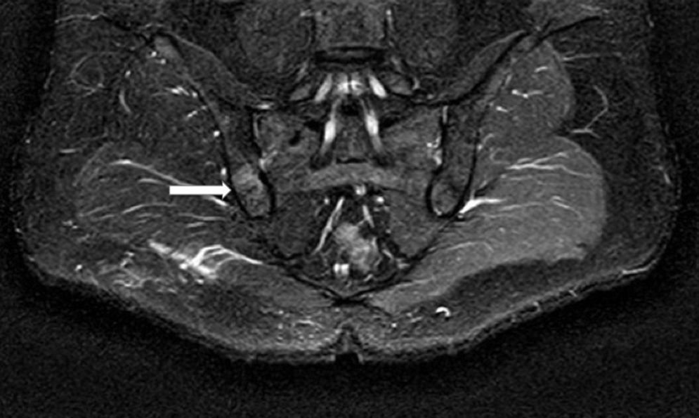
The patient complained of pain in right buttock that waked him up during the night and was relieved by nonsteroidal anti-inflammatory drugs (NSAID). He had no history of traumatic injury, fever or arthritis, and reported nothing during work-up that suggested spondyloarthritis or recent infections. The physical examination was completely normal, with negative results in sacroiliac pain provocation tests (flexion, abduction, external rotation [FABER], distraction, compression and Laguerre tests), and the joint showed no peripheral or axial functional limitations. The neurological examination revealed no evidence of disease. The results of the laboratory tests, including the acute phase reactant levels, were normal. With respect to imaging studies, as there were no evident bone changes on plain radiography of the pelvis, the patient underwent magnetic resonance imaging (MRI) of the sacroiliac joints. There was a hypointense area on T1-weighted sequences (Fig. 1) and a hyperintense area in short tau inversion recovery (STIR) images (Fig. 2) on the border of the iliac side of the right sacroiliac joint, with an isointense central zone on T1-weighted sequences (Fig. 1), which was hypointense on the STIR sequences (Fig. 2). These findings pointed toward a definitive diagnosis of osteoid osteoma, which was confirmed by computed tomography (CT). The patient was treated immediately by means of CT-guided radiofrequency ablation and his symptoms disappeared.
Osteoid osteoma is a benign bone tumor that occurs in the femur or tibia in 50%–60% of the cases; between 7% and 10% are located in the spine.1 Pelvic osteoid osteoma, like that of our patient, is less common. It occurs more frequently in males between the ages of 10 and 30 years. A typical symptom is nocturnal pain, which is relieved by NSAID.2 The pattern of nighttime pain associated with this type of tumor can lead to its being mistaken, as in the case we report, for certain inflammatory rheumatic diseases, especially when located at certain sites, a circumstance that can result in a delay in the diagnosis. Although the osteoid osteoma is not often included in the differential diagnosis of sacroiliac pain, we should take it into account when the pain is relieved by NSAID, and in the case of young patients in whom the pain does not respond to conventional treatment.3
Plain radiography does not always show the typical image with the nidus. Thus, it may be necessary to resort to other techniques such as MRI and/or CT (as MRI may not be very helpful, CT is the technique of choice in this type of tumor), since bone scintigraphy has a high sensitivity, but a low specificity.2 Computed tomography-guided radiofrequency ablation is the preferred therapeutic approach, as it is less invasive than other techniques and the outcome is good.1
Please cite this article as: Moreno-Martinez MJ, Moreno-Ramos MJ, Díaz-Navarro MJ, Linares-Ferrando LF. Osteoma osteoide pélvico simulando sacroileitis. Reumatol Clin. 2016;12:177–178.


