


Autoimmune hepatitis (AIH) is a chronic inflammatory liver disease with low prevalence worldwide. The coexistence of this entity with rheumatic diseases has been evaluated in multiple studies and is highly variable. The objective of this study is to identify the frequency of coexistence of rheumatic diseases and autoimmune hepatitis in adults who have been treated for 10 years in a fourth-level hospital in Bogota, Colombia.
Materials and methodsAnalytical, observational, cross-sectional study in a single center that included patients over 18 years of age of both sexes with a diagnosis of AIH by simplified score ≥7 points, with a medical history registered at the Fundacion Santa Fe de Bogota in Bogota, Colombia between January 2013 and December 2023.
ResultsA total of 66 patients met inclusion criteria. 36.4% of patients had a concomitant autoimmune disease, with Sjögren's syndrome, systemic lupus erythematosus and autoimmune thyroid disease being the most prevalent.
ConclusionThe frequency of coexistence of autoimmune hepatitis with rheumatic diseases in adult patients is 36.4% for the cohort studied, which is within the range of what has already been reported globally, where a prevalence of 14 to 44% has been described.
La hepatitis autoinmune (HAI) es una enfermedad crónica inflamatoria del hígado de baja prevalencia a nivel global. En múltiples estudios se ha evaluado la coexistencia de esta entidad con enfermedades reumatológicas la cual es muy variable. El objetivo de este estudio es identificar la frecuencia de la coexistencia de enfermedades reumatológicas y hepatitis autoinmune en adultos que han sido atendidos durante 10 años en un hospital de cuarto nivel en Bogotá, Colombia.
Materiales y métodosEstudio observacional analítico de corte transversal en un único centro que incluyó pacientes mayores de 18 años de ambos sexos con diagnóstico de HAI por “score” simplificado ≥7 puntos, con historia clínica registrada en el Hospital Fundación Santa Fe de Bogotá entre enero de 2013 y diciembre 2023.
ResultadosUn total de 66 pacientes cumplieron criterios de inclusión. El 36.4% de los pacientes presentaban una enfermedad autoinmune concomitante, siendo el síndrome de Sjögren, el lupus eritematoso sistémico y la enfermedad tiroidea autoinmune las de mayor prevalencia.
ConclusiónLa frecuencia de la coexistencia de hepatitis autoinmune con enfermedades reumatológicas en pacientes adultos es del 36.4% para la cohorte estudiada, encontrándose en el rango de lo ya reportado a nivel global, donde se ha descrito una prevalencia del 14 al 44%.
Autoimmune hepatitis (AIH) is a rare chronic inflammatory liver disease that follows an erratic course and characterised by elevated transaminases, hypergammaglobulinaemia, the presence of antibodies, and typical histopathological lesions.1 It has an incidence of 1.37 per 100,000 inhabitants and a prevalence of 17.4 per 100,000 inhabitants worldwide.2
Numerous studies performed around the world have evaluated the coexistence of this condition with rheumatological diseases, reporting prevalence rates that vary from 28% to 40%, which speaks to a high degree of variability owing to the heterogeneity of the cohorts studied. Ascertaining the frequency with which rheumatological diseases co-exist in AIH in our setting is pertinent given the impact they have on the natural history of the disease, treatment, and prognosis, bearing in mind that if comorbidities are diagnosed early, they can affect the patient's quality of life.3
The epidemiology of AIH has not been fully elucidated. Depending on the cohort studied, incidences of 1-2 per 100,000 person-years in adult patients and one-off prevalence rates of 11-17 per 100,000 person-years in Norway and Sweden have been reported.4 Alaska reports a particularly high prevalence of the disease (43 per 100,000), with a significant proportion of cases debuting with jaundice.5 One of the obstacles that hinder access to reliable epidemiological data regarding AIH is that some studies include individuals diagnosed without histological confirmation and/ or scoring systems or subjects with other aetiologies of liver disease, such as MAFLD and viral hepatitis with the presence of autoantibodies.6
AIH can debut at any age; however, two peaks of incidence have been documented; specifically, between 10 and 30 years and 40 and 60 years of age.7 It afflicts all ethnicities and Black patients with AIH are reported to be more likely to have an earlier age of onset and liver failure with a greater risk of requiring liver transplantation despite experiencing a comparable response to steroids as non-Black individuals.8
The evidence currently available on the subject is meagre and many questions remain unanswered, which is why this research seeks to identify how often AIH coexists with rheumatological diseases in a fourth-level hospital in Bogota, Colombia. An observational, analytical, retrospective study was carried out by reviewing the medical records of patients with AIH over a 10-year period, in which a cohort of the population with AIH and concomitant autoimmune diseases was obtained. We conducted a sociodemographic characterisation of this population, as well as an analysis of their clinical variables and, more specifically, their immune-serological profile. As for the limitations of this study, of the 616 case histories reviewed, only 66 patients met the inclusion criteria for analysis, and the vast majority of them did not have a complete immune-serological profile.
MethodologyType of studySingle-centre, cross-sectional, analytical observational study.
Population and sampleA data base was created containing all those patients who consulted the Fundación Santa Fe de Bogotá between 2013 and 2023 and who had a diagnosis of AIH according to the ICD-10 classification, coded as K75.4. Once this data base had been constructed, all data pertaining to repeat cases were purged in order to obtain the total number of subjects with an ICD-10 K75.4 diagnosis of AIH who had consulted the institution during that period.
Afterwards, the diagnostic criteria of the AIH score were applied, so as to confirm those patients who actually had a diagnosis of AIH. These diagnostic criteria were calculated using the earliest values available in the registry, following the result of the liver biopsy. Once the inclusion criteria had been applied to the study, including the AIH score ≥ 7 points, as well as the exclusion criteria, the data were collected in the data base.
Sociodemographic variablesPatient age and sex were recorded.
Clinical characteristicsThe presence of cirrhosis, the Child-Pugh classification, and the existence of overlap syndrome were recorded. Similarly, relevant paraclinical variables were also identified and logged: liver profile and function, platelet count, immune-serological profile of autoimmune liver disease.
Furthermore, the existence of extrahepatic autoimmune disease and the associated immune-serological profile were also noted. In other words, the diagnosis of autoimmune illness by disease-related background in the clinical history could include: systemic lupus erythematosus, rheumatoid arthritis, Sjögren´s syndrome, systemic sclerosis, systemic vasculitis, inflammatory myopathy, mixed connective tissue disease, autoimmune thyroid disease (Graves' disease, Hashimoto's thyroiditis), inflammatory bowel disease (Crohn's disease, ulcerative colitis), polymyalgia rheumatica, fibromyalgia, sarcoidosis, psoriasis, vitiligo, diabetes mellitus type 1, multiple sclerosis, celiac disease, antiphospholipid syndrome. On the other hand, the following antibodies will be included in the immune-serological profile: anti-DNA, anti-RO, anti-La, anti-RNP, anti-SM, anti-CCP, rheumatoid factor, P-ANCA, C-ANCA, anti-MPO, anti-PR3, anti-SCL-70, anticentromere.
Finally, the histological findings in those subjects with a liver biopsy pathology report were reported.
Sample sizeGiven the observational nature of the study, all those cases meeting the inclusion and exclusion criteria in the study timeline (January 2013-20223) were admitted.
Inclusion criteria- •
Patients over the age of 18 years who present at hospitalisation, the emergency room, or outpatient clinic belonging to the Fundación Santa Fe de Bogotá between 2013 and 2023 with a diagnosis of AIH based on the simplified AIH score of 7 points or more.
- •
Complete data: Patients whose medical history contains full information regarding their clinical and demographic, as well as their autoimmune profile information (Table 1).
Table 1.Simplified autoimmune hepatitis diagnostic criteria.
Variable Cutoff Score ANA or SMA >1:40 1 ANA or SMA >1:80 2a Anti-LKM >1:40 2a Anti-SLA >1:40 2 Serum IgG >Upper limit of normal 1 Serum IgG >1.1 times the upper limit of normal 2 Liver histology Compatibleb 1 Liver histology Typicalc 2 Absence of viral hepatitis Yes 2
- •
Patients under the age of 18 years.
- •
Incomplete AIH.
- •
Simplified AIH score below 7.
Because of the descriptive, observational nature of the study, no hypothesis was put forward with respect to the frequency of rheumatological diseases coexisting with AIH.
Data gathering techniqueA data base was used as the tool with which to record and compile the information. The information was collected by reviewing the medical records of previously identified patients with a diagnosis of AIH as per ICD-10 coding and stored in a data base by means of the REDCAP application as the only means authorised by the Fundación Santa Fe de Bogotá to log and analyse the data. They were then meticulously integrated into an Excel spreadsheet, with prior anonymisation of the information to guarantee the participants' privacy and confidentiality. Since, for the purposes of this study, the identification number is the only value that can be directly used to identify a person, once all the information concerning the study had been collected, these data were eliminated and a numerical value was assigned that did not make it possible to identify the subjects at a later date.
Statistical aspectsThis is a descriptive analytical, observational, cross-sectional study in which an analysis of the socio-demographic description was made, and qualitative variables were determined in the form of absolute and relative frequency and percentages with their respective confidence intervals. Furthermore, quantitative variables were established by means of measures of central tendency, dispersion, and 95% confidence intervals, with the mean and standard deviation for those exhibiting normal distribution, and median and interquartile range for those displaying a non-normal distribution.
Possible associations were ascertained using the chi-square statistic or ANOVA, depending on the type of variable, taking into account a significance level of 5% (p < 0.05). All the analyses described above were made with the statistical software IBM SPSS Statistics version 29.0.10.
Informed consentGiven that, according to article 11 of resolution 8430 of 1993, this research has been regarded as “risk-free”, informed consent was not mandatory.
ResultsA total of 616 patients were identified as presenting AIH with ICD-10 diagnosis K75.4 between 2013 and 2023. Out of these 616 cases, only 66 patients met the inclusion criteria for further analysis of the study data (Fig. 1).

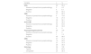
Ninety-seven percent of the subjects were female and only two individuals were male. The mean age of the sample was 57 years, with a range of 29 to 82 years at the time of diagnosis of AIH (Table 2).
Cirrhosis was present in 84.8% of the patients examined at the time of diagnosis, with Child-Pugh classification A and B being the primary classification, accounting for 48% and 41%, respectively. In contrast, 8% of the patients presented Child-Pugh C (Table 3). Among the paraclinical parameters, transaminases were observed with mean values of 165 UI/l and 150 UI/l for AST and ALT, respectively, and mean alkaline phosphatase was 312 UI/l, and [mean] GGT was found to be 343 UI/l. The paraclinical indicators of liver function included an albumin of 3.4 mg/dl; prothrombin time was not indicative of INR prolongation, platelet counts were within normal ranges, and direct bilirubin of 2.1 and indirect bilirubin averaged 1.3 (Table 4).
Clinical characteristics of patients with AIH (n = 66).
| Variable | Minimum | Maximum | Mean |
|---|---|---|---|
| AST | 19 | 1614 | 165 |
| ALT | 15 | 1440 | 150 |
| Direct bilirubin | 0.11 | 18.37 | 2.19 |
| Indirect bilirubin | 0.14 | 10.65 | 1.38 |
| Alkaline phosphatase | 55 | 1454 | 312 |
| GGT | 16 | 2610 | 343 |
| Prothrombin time | 9.46 | 61 | 13.6 |
| Albumin | 1.5 | 4.6 | 3.46 |
| Platelets | 62,000 | 469,000 | 215,000 |
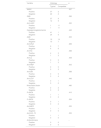
Insofar as the variables considered as part of the simplified diagnostic criteria for AIH are concerned, 63% of the patients analysed had the typical histopathological findings of AIH, while the remaining 36% presented findings that were consistent with AIH (Table 5). As for the immunoserological profile, 89.4% of the subjects tested positive for antinuclear antibodies; 59.1% were found to have anti-smooth muscle antibodies, and 98.1% displayed hypergammaglobulinaemia. Moreover, 42% of cases had no paraclinical tests for liver and kidney microsomal 1 antibodies available, with only 3% presenting these antibodies, and 39.4% of patients with anti-mitochondrial antibodies (Table 5). Finally, 43.9% of the sample displayed overlap syndrome with primary biliary cirrhosis.
Hepatic autoimmune profile and histopathological findings (n = 66).
| Variable | n | % |
|---|---|---|
| ANA | 63 | 95.4 |
| Absence of paraclinical symptomatology | 3 | 4.5 |
| Negative | 4 | 6.1 |
| Positive | 59 | 89.4 |
| SMA | 57 | 86.3 |
| Absence of paraclinical symptomatology | 9 | 13.6 |
| Negative | 18 | 27.3 |
| Positive | 39 | 59.1 |
| Anti-LKM1 | 24 | 36.3 |
| Absence of paraclinical symptomatology | 42 | 63.6 |
| Negative | 22 | 33.3 |
| Positive | 2 | 3 |
| Hypergammaglobulinemia | 66 | 100 |
| Absence of paraclinical symptomatology | 0 | 0 |
| Negative | 1 | 1.5 |
| Positive | 65 | 98.5 |
| AMA | 47 | 71.2 |
| Absence of paraclinical symptomatology | 19 | 28.8 |
| Negative | 21 | 31.8 |
| Positive | 26 | 39.4 |
| Histology | 66 | 100 |
| Typical | 42 | 63.6 |
| Compatible | 24 | 36.4 |
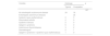
Of the 66 individuals studied, 36.4% had an associated extrahepatic autoimmune disease, with Sjögren’s syndrome being the most prevalent, followed by autoimmune thyroid disease and systemic lupus erythematosus (Table 6). The autoimmune profile of the antibodies that were evaluated was positive for anti-DNA, anti-Ro, anti-La, rheumatoid factor, P-ANCA, MPO, and anti-SCL-70; anti-DNA and anti-Ro were the most prevalent (Table 7).
Types of autoimmune disease in patients with autoimmune hepatitis (n = 66).
| Variable | n | % |
|---|---|---|
| No extrahepatic autoimmune disease | 42 | 63.6 |
| Systemic lupus erythematosus | 5 | 7.1 |
| Rheumatoid arthritis | 2 | 3 |
| Systemic sclerosis | 2 | 3 |
| Sjögren’s syndrome | 6 | 9.1 |
| Autoimmune thyroid disease | 5 | 7.6 |
| Dermatological | 3 | 4.5 |
| Sjögren’s syndrome + systemic lupus erythematosus | 1 | 1.5 |
Statistical analysis for the association between histology and antibodies.
| Variable | Histology | p | |
|---|---|---|---|
| Typical | Compatible | ||
| ANAS | .357 | ||
| Positive | 37 | 2 | |
| Negative | 22 | 2 | |
| SMA | .366 | ||
| Positive | 27 | 9 | |
| Negative | 12 | 9 | |
| Anti-LKM1 | .220 | ||
| Positive | 1 | 11 | |
| Negative | 1 | 11 | |
| Hypergammaglobulinemia | .446 | ||
| Positive | 41 | 1 | |
| Negative | 24 | 0 | |
| AMA | .935 | ||
| Positive | 16 | 14 | |
| Negative | 10 | 7 | |
| Anti-DNA | .689 | ||
| Positive | 9 | 7 | |
| Negative | 4 | 6 | |
| Anti-Ro | .588 | ||
| Positive | 4 | 3 | |
| Negative | 1 | 3 | |
| Anti-La | .408 | ||
| Positive | 0 | 5 | |
| Negative | 1 | 3 | |
| Anti-RNP | .943 | ||
| Positive | 0 | 5 | |
| Negative | 0 | 3 | |
| Anti-SM | .588 | ||
| Positive | 0 | 5 | |
| Negative | 0 | 4 | |
| Anti CCP | .446 | ||
| Positive | 0 | 1 | |
| Negative | 0 | 0 | |
| Rheumatoid factor | .480 | ||
| Positive | 2 | 2 | |
| Negative | 0 | 2 | |
| P-ANCA | .410 | ||
| Positive | 1 | 1 | |
| Negative | 0 | 2 | |
| C-ANCA | .264 | ||
| Positive | 0 | 1 | |
| Negative | 0 | 2 | |
| Anti-MPO | .555 | ||
| Positive | 1 | 1 | |
| Negative | 0 | 0 | |
| Anti-SCL-70 | .555 | ||
| Positive | 1 | 1 | |
| Negative | 0 | 0 | |
| Anticentromere | – | ||
| Positive | 0 | 0 | |
| Negative | 0 | 0 | |
Statistical analyses were carried out to investigate possible associations between histopathological findings and the extrahepatic autoimmune profile; no such associations were detected (Table 7). Similarly, no associations were found between histopathological findings and extrahepatic autoimmune diseases (Table 8). We also found no association between the different types of autoimmune disease and the presence of cirrhosis (Table 9).
Statistical analysis for the association between histology and type of autoimmune disease.
| Variable | Histology | p | |
|---|---|---|---|
| Typical | Compatible | ||
| .307 | |||
| No extrahepatic autoimmune disease | 24 | 18 | |
| Extrahepatic autoimmune disease | 22 | ||
| Systemic lupus erythematosus | 2 | 3 | |
| Rheumatoid arthritis | 2 | 0 | |
| Systemic sclerosis | 2 | 0 | |
| Sjögren’s syndrome | 6 | 0 | |
| Autoimmune thyroid disease | 3 | 2 | |
| Dermatological | 2 | 1 | |
| Sjögren’s syndrome + Systemic lupus erythematosus | 1 | 0 | |
Statistical analysis for the association between cirrhosis and type of autoimmune disease.
| Variable | Cirrhosis | p | |
|---|---|---|---|
| No | Yes | ||
| .589 | |||
| No extrahepatic autoimmune disease | 8 | 34 | |
| Extrahepatic autoimmune disease | 2 | 22 | |
| Systemic lupus erythematosus | 1 | 4 | |
| Rheumatoid arthritis | 1 | 1 | |
| Systemic sclerosis | 0 | 2 | |
| Sjögren’s syndrome | 0 | 6 | |
| Autoimmune thyroid disease | 0 | 5 | |
A total of 616 patient records with an ICD-10 K75.4 diagnosis of AIH were identified within the 10 year period studied. Of them, though, 62% could not be analysed for the reasons detailed in Fig. 1. It is worth noting that 109 of these records had no liver histology results available, despite the fact that they had a diagnosis of AIH. This suggests that in clinical practice the diagnostic score is not applied to define the presence of this disorder. There were 95 patient records of individuals who were coded with the ICD-10 diagnosis of AIH but who, following extension studies, were found to actually have another condition. In addition, 58 clinical records were located that, following a comprehensive review and after ICD-10 diagnosis, were incompatible in terms of clinical, paraclinical, and histological parameters, and therefore failed to fulfil the criteria for the AIH score; nevertheless, these patients were classified as having AIH, and were therefore not taken into account in the final analysis. This may reveal that while the study was performed with individuals who were evaluated in a referral centre for liver transplantation in Colombia, diagnosing this condition poses a challenge for the specialist.
A higher prevalence of the disorder was detected in women, as well as an average age of 57 years, which is in line with prevalence rates reported around the world.2,4 Five individuals were found who were 75 years old or more, which points to the fact that this diagnosis should not be ruled out at these extremes of age when clinical and paraclinical findings are suggestive of the disease.
Of the cohort that underwent analysis, 84.8% had cirrhosis at diagnosis; 48% exhibited compensated cirrhosis (Child A), and 49% were decompensated (Child B and C). This result contrasts markedly with the observations made by Díaz-Ramírez et al. in their study9 in which only 17% presented cirrhosis at diagnosis and none were Child C. We believe this difference may be due to the facility where the study was carried out inasmuch as it is a referral centre for hepatology, it may receive people whose condition is more advanced than at other institutions.
As regards the histopathological findings, which in this study were classified as typical and consistent on the basis of the simplified AIH score, very similar data were recorded in terms of distribution as those in the work carried out in Cali by Díaz-Ramírez et al.9 This indicates that the histopathological behaviour of the disease is identical in both cohorts under study and may conceivably be extrapolated to the rest of the country.
Based on the results of the autoimmune profile aimed at the diagnosis of AIH using a simplified score, by and large, the diagnosis was made according to ANA positivity, anti-SMA, and hypergammaglobulinaemia. It is important to highlight the scant use of anti-LKM1 in clinical practice, which may be indicative of an underreporting of type 2 AIH in our population.
A 43.9% prevalence rate of overlap with PBC was detected in our cases; although, this diagnosis was not confirmed when the Paris criteria were considered. In comparison to international works, this could potentially reflect overdiagnosis, given that the prevalence rates reported in the literature ranged from 2 to 19%.10
With respect to concomitant autoimmune disease, 24 of our 66 subjects evidenced some autoimmune disease, primarily Sjögren’s syndrome (25%), systemic lupus erythematosus (20.8%), and autoimmune thyroid disease (20.8%). Only a single case of Sjögren’s syndrome with systemic lupus erythematosus is reported in a patient with AIH. These findings are in line with other studies in Latin America, such as the one by Paredes Millán et al.11 in Lima, Peru, as well as the one performed by Díaz-Ramírez et al.9 in Cali, Colombia.9
If we look at the extrahepatic autoimmune serological profile, we find that the vast majority of these individuals do not have this type of study available to them, which indicates that they were requested when a particular condition was suspected, not as a screening tool for any other autoimmune disease. While a higher prevalence of anti-DNA and anti-Ro positivity is evidenced, the statistical analysis for associations with histopathological findings found no correlation between these variables, which would imply that the current practice of requesting these studies when rheumatological disease is suspected should be maintained. Likewise, we sought out associations between the type of extrahepatic autoimmune disease and histopathological findings; this yielded a p-value of 0.307, indicating no statistically significant association. This implies that histopathological findings do not depend on whether there is a greater or lesser burden of systemic autoimmunity. In the same way, no relationship was apparent between the type of autoimmune disease and the presence of cirrhosis at the time of AIH diagnosis. It is unclear whether concomitant autoimmune disease results in worse outcomes for AIH in terms of progression to fibrosis and complications from portal hypertension. Nonetheless, having rheumatological comorbidity increases morbidity and diminishes the patient's quality of life.3 In this study, we did not examine whether concomitance increased the risk of progression to cirrhosis and its complications or if it entailed any prognostic implications. Further studies will be needed to answer these questions.
Methodologically speaking, given the nature of the study conducted and for one reason or another, some people who have a diagnosis of AIH were not coded with the ICD-10 code K75.4 during the study period and were therefore excluded from consideration. Moreover, the simplified score was chosen as the diagnostic criterion for AIH, which, though it has greater specificity and accuracy than the revised IAIHG score, the latter is more sensitive for cases with unusual or more complex presentations, which may have resulted in patients being excluded who would, in fact, have met the diagnostic criteria as per the revised score.6,12 Furthermore, there is a percentage of patients who may be seronegative for AIH depending on how they present (<7% who debut in acute forms and between 1% and 34% in chronic forms12) and who, because of our inclusion criteria, were not included in the study. Another limitation derives from information bias, in view of the retrospective nature of the study and the review of medical records, i.e., 103 individuals with missing data that might have represented a larger sample to be analysed in the study were excluded because of the meticulous review of the medical records.
It is still worth noting the previously mentioned overlap syndrome with PBC, in which the Paris criteria to confirm the diagnosis of overlap syndrome were not verified, which may point to possible overdiagnosis compared to reports in the literature.
It should also be mentioned that despite the fact that this is a single-centre study, it is a referral centre for liver disease in Colombia, and the results previously mentioned are very much in line with what has been found in the literature, which lends some measure of external validity to the study.
One of the strengths of the study is that, to the best of our knowledge, this is only the second study in Colombia to characterise patients with AIH, which is important because it contributes to what is known about this condition in our region. Furthermore, this is the first time that the extrahepatic autoimmune profile of these people has been described in Colombia. More specifically, this is the first study in the literature to explore the association between histopathological findings and the presence of cirrhosis at diagnosis in individuals who present AIH and concomitant extrahepatic autoimmune diseases.
ConclusionsIn summary, the frequency with which AIH coexists with rheumatological diseases in adult patients is 36.4% for the cohort studied, which is in the range of previously reported global prevalence rates of 14-44%.13 Of the sociodemographic characteristics studied, the predominant sex and average age at presentation are similar to reports in the literature from around the world. Nevertheless, unlike other cohorts, a higher prevalence of cirrhosis was detected at the time of presentation, in more advanced stages of decompensation, which is probably related to the fact that our work was performed in a referral centre for hepatology. On the other hand, in spite of not finding any connection between the type of extrahepatic autoimmune diseases and the immunoserological profile with the presence of cirrhosis or histopathological findings, this is the first time this type of association has been examined, which therefore provides novel information and confirms that an immunoserological study should not be routinely performed in all cases of AIH unless a particular rheumatological entity is suspected. We encourage further efforts to search for other possible associations that may have an impact on the prognosis and survival of patients with AIH.
The authors have no conflict of interests to declare.


