Journal Information
Vol. 15. Issue 3.
Pages 182-184 (May - June 2019)
Vol. 15. Issue 3.
Pages 182-184 (May - June 2019)
Images in Clinical Rheumatology
Full text access
Unusual Clinical Complication: Acute Lower Limb Ischemia Caused by a Tibial Osteochondroma
Complicación clínica inusual: isquemia aguda de miembro inferior causada por osteocondroma tibial
Visits
3617
Noelia Alonso-Gómeza,
Corresponding author
nelinew@hotmail.com

Corresponding author.
, Miguel Cuesta-de Diegob, Antonio Martínez-Izquierdoa, Felipe Sáinz-Gonzáleza
a Servicio de Angiología y Cirugía Vascular, Hospital Universitario Central de la Defensa Gómez-Ulla, Madrid, Spain
b Unidad Médica Aérea de Apoyo al Despliegue del Ejército del Aire (UMAAD), Madrid, Spain
This item has received
Article information
Full Text
Bibliography
Download PDF
Statistics
Figures (4)
Show moreShow less
Full Text

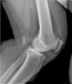
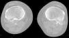
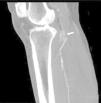
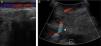
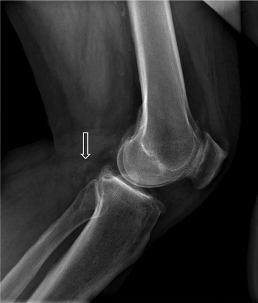
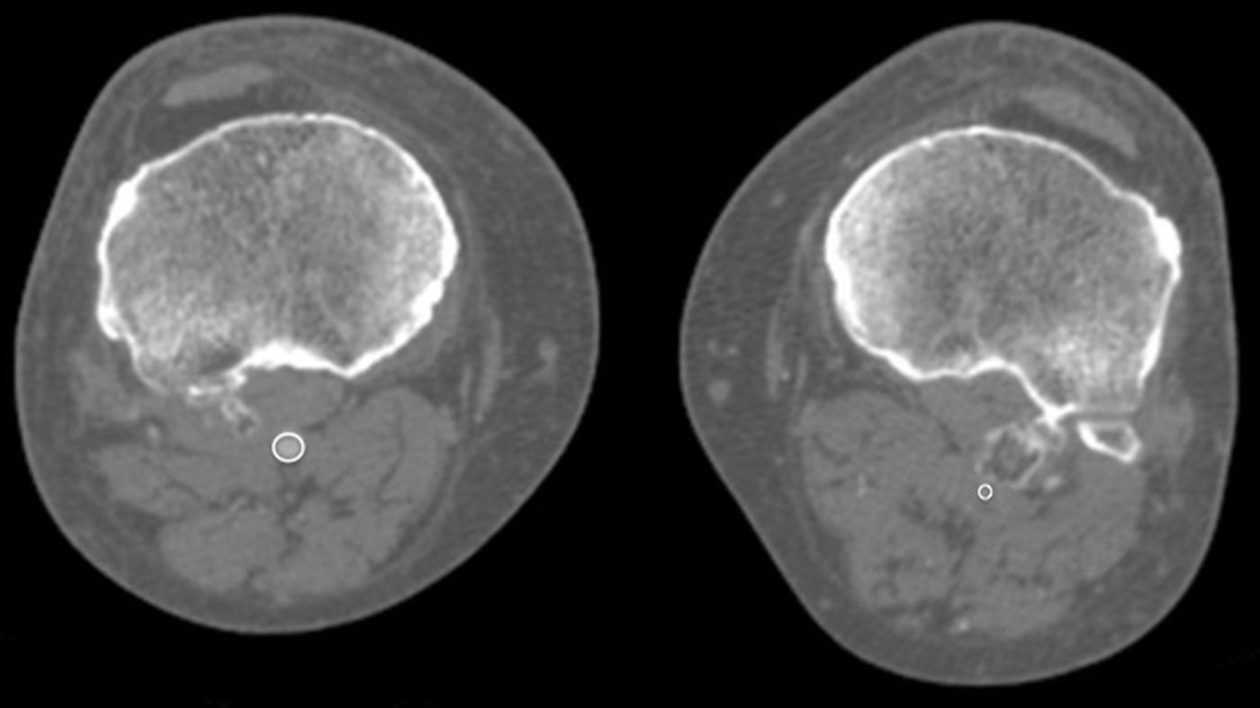
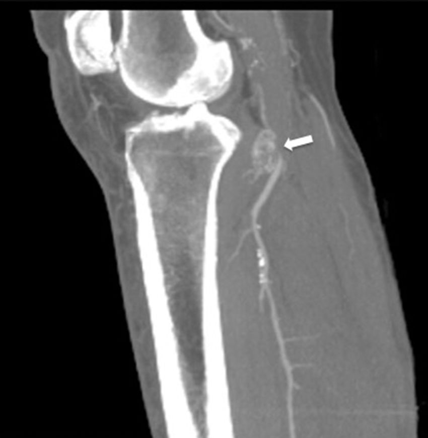
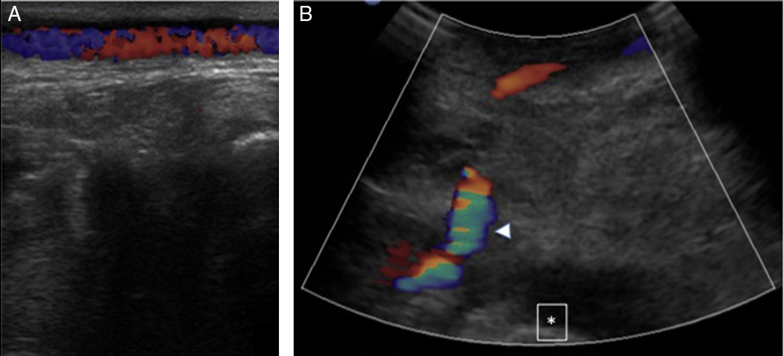
A male, 67 years of age, obese, with high blood pressure and dyslipidemia presented at the emergency department due to pain, cold and functional impairment of his lower left limb of 24-h onset. During anamnesis, the patient stated he had had a chronic intermittent pain in his left knee for a long time. A previous simple knee X-ray showed an osteochondroma (Fig. 1). The patient presented with a femoral pulse but with no popliteal and distal pulses. Artery examination was normal in the contralateral extremity. A CT angiogram was requested for the lower limbs which showed thrombosis of the popliteal artery adjacent to the osteochondroma (Figs. 2 and 3). We decided to perform in situ revascularization with femoro-popliteal saphenous vein graft (Fig. 4) avoiding the tunneling technique for anatomical positioning. To date, after 3 years of follow-up with eco-Doppler, the bypass continues maintaining permeability with ankle-arm index of 0.9, and no signs of popliteal vein injury.

Figure 1.

Lateral X-ray of the knee: exophytic bone lesion (arrow) tibia-dependent corresponding to an epiphyseal osteochondroma.

(0.05MB).
Figure 2.

CT angiogram of MMII. Axial slice. Posterior displacement of the left popliteal artery (circles).

(0.06MB).
Figure 3.

CT angiogram. Sagittal slice. Maximum intensity projection (MIP). Thrombosis of the popliteal artery (arrow) with distal rechanneling.

(0.03MB).
Figure 4.

Eco-Doppler: bypass control. (A) Superficial pathway of the in situ SVG. (B) Permeable bypass (arrow head). Femoral condyle (asterisk).

(0.09MB).

Osteochondroma is the most common pseudotumoral bone lesion. The radiologic pathognomonic characteristic of this tumor is the cortical and medullar continuity of the lesions with the bone from which they protrude.1 They may be single or multiple.2 Some of the most common complications are the presence of bone deformity, fractures,1 vascular compromise3 and neurological compromise,4 formation of a bursa5 and malignant degeneration.6 Diagnosis may be made with an X-ray, although other imaging methods such as a scan, CT and MR may be used in suspected cases when symptoms occur or are in unusual locations.1 Vascular compromise may be caused by displacement of blood vessels (arteries and veins), by stenosis, occlusion and the formation of pseudoaneurisms, with the latter being more frequent at knee level,7 involving the popliteal artery or being presented as arterial thrombosis,8,9 as in this clinical case. Its presentation as thoracic outlet syndrome in the case of rib lesions has been described.10

Familiarity with the spectrum of radiological findings leads to an accurate diagnosis which is helpful in managing the patient toward appropriate treatment.

Ethical LiabilitiesProtection of people and animals

The authors declare that no experiments using human beings or animals have been carried out for this research study.

Data confidentiality

The authors declare that they have followed the protocols of their center of work on patient data publication.

Right to privacy and informed consent

The authors declare that they obtained the informed consent from the patients and/or subjects referred to in this article.

Conflicts of Interest

The authors have no conflicts of interest to declare.

References
[1]
M.D. Murphey, J.J. Choi, M.J. Kransdorf, D.J. Flemming, F.H. Gannon.
Imaging of osteochondroma: variants and complications with radiologic–pathologic correlation.
Radiographics, 20 (2000), pp. 1407-1434
[2]
J.V. Bovée.
Multiple osteochondromas.
Orphanet J Rare Dis, 3 (2008), pp. 3
[3]
K. Gruber-Szydło, R. Poręba, K. Belowska-Bień, A. Derkacz, R. Badowski, R. Andrzejak, et al.
Popliteal artery thrombosis secondary to a tibial osteochondroma.
[4]
A. Turan Ilica, E. Yasar, H. Tuba Sanal, C. Duran, I. Guvenc.
Sciatic nerve compression due to femoral neck osteochondroma: MDCT and MR findings.
Clin Rheumatol, 27 (2008), pp. 403-404
[5]
S.M. Mahmoodi, R.K. Bahirwani, B.A. Abdull-Gaffar, I.F. Habib.
Intrabursal vein abrasion and thrombosis. an unusual complication of femoral osteochondroma.
Saudi Med J, 30 (2009), pp. 1604-1606
[6]
C. Martin, P.L. Munk, J.X. O’Connell, M.J. Lee, B. Masri, N. Wambeek.
Malignant degeneration of an osteochondroma with unusual intra-bursal invasion.
Skeletal Radiol, 28 (1999), pp. 540-543
[7]
F. Raherinantenaina, H.N. Rakoto-Ratsimba, T.M. Rajaonanahary.
Management of extremity arterial pseudoaneurysms associated with osteochondromas.
Vascular, 24 (2016), pp. 628-637
[8]
I. Khan, C.A. West Jr., G.P. Sangster, M. Heldmann, L. Doucet, M. Olmedo.
Multiple hereditary exostoses as a rare nonatherosclerotic etiology of chronic lower extremity ischemia.
J Vasc Surg, 51 (2010), pp. 1003-1005
[9]
N. Tanigawa, S. Kariya, H. Kojima, A. Komemushi, H. Fujii, S. Sawada.
Lower limb ischaemia caused by fractured osteochondroma of the femur.
Br J Radiol, 80 (2007), pp. e78-e80
[10]
P.J. O’Brien, S. Ramasunder, M.W. Cox.
Venous thoracic outlet syndrome secondary to first rib osteochondroma in a pediatric patient.
J Vasc Surg, 53 (2011), pp. 811-813

Please cite this article as: Alonso-Gómez N, Cuesta-de Diego M, Martínez-Izquierdo A, Sáinz-González F. Complicación clínica inusual: isquemia aguda de miembro inferior causada por osteocondroma tibial. Reumatol Clin. 2019;15:182–184.

Copyright © 2017. Elsevier España, S.L.U. and Sociedad Española de Reumatología y Colegio Mexicano de Reumatología
Idiomas
Reumatología Clínica (English Edition)
Article options
Tools
es en

¿Es usted profesional sanitario apto para prescribir o dispensar medicamentos?

Are you a health professional able to prescribe or dispense drugs?