


There are many factors that influence the pathogenesis of autoimmune disease of which host genetic factors play an important role. The aim of this study was to investigate the HLA Class I and II genes in a family with a high incidence of AID to establish whether they contribute to the development of these disease.
Four of them had been diagnosed with SLE and one with AHA. The patients with SLE showed the presence of HLA-A*02 B*40 DRB1*04:07 DQB1*03:02 haplotype with a high statistical significance. This haplotype was not present in the healthy individuals and in the patient with AHA, although the DRB1*04:07 DQB1*03:02 haplotype (carried by both parents) was found in the AHA patients and one of the healthy individuals.
We must consider how HLA Class I in linkage disequilibrium with HLA Class II may be involved in susceptibility or in the development of SLE. An extensive study in this population should be conducted to establish the true participation of the HLA Class I region.
Muchos factores se han involucrado en la patogénesis de las enfermedades autoinmunitarias, entre los cuales el fondo genético desempeña un papel importante. El objetivo fue investigar los genes de HLA Clase I y II en una familia con alta incidencia de enfermedades autoinmunitarias para establecer si estos podrían contribuir al desarrollo de estas enfermedades.
Los pacientes diagnosticados de lupus mostraron la presencia del haplotipo HLA A*02, B*40, DRB1*04:07, DQB1*03:02 con alta significación estadística. En los individuos sanos y en el paciente con AHA este haplotipo no estuvo presente; en cambio, el haplotipo Clase II DRB1*04:07, DQB1*03:02, estuvo también en el paciente con AHA y en uno de los individuos sanos.
Deberíamos considerar cómo HLA Clase I en desequilibrio de ligamiento con HLA Clase II podría estar involucrado en la susceptibilidad o el desarrollo de lupus eritematoso sistémico. Un estudio más extenso de esta población debería llevarse a cabo a fin de establecer la verdadera participación de la región de HLA Clase I.
Pathogenesis of autoimmune disease (AID) is associated with functional deficiency of multiples immunologic component including innate immune system. The failure of mechanisms regulatory could be due to the influence of multiple genetic factors to contribute to disease susceptibility.1,2
The search for genes in systemic lupus erythematosus (SLE) have focused, on the highly polymorphic HLA Class I and II genes as well as genes within the HLA Class III region, particularly tumor necrosis factor (TNF) and complement component C4 gene loci.
Work by Graham et al.3 involving an analysis of approximately 50 microsatellite genetic markers across the HLA region among a set of 780 SLE families, the importance of HLA Class II haplotypes involving the HLA-DRB1 and -DQB1 loci, particularly those corresponding to serologic types HLA-DR2 and DR3.
GWAS (genomic-wide association studies) in both European and Asian populations have shown that the strongest contribution to risk for SLE resides in the HLA region have identified more than 30 associated including genetic variants of HLA and Fcγ receptor genes, IRF5, STAT4, PTPN22, TNFAIP3, BLK, BANK1, TNFSF4 and ITGAM. However, these loci account for less than 10% of the genetic heritability.3–5
Population studies reveal that the susceptibility to SLE involves human leukocytes antigen (HLA) Class II. An association of HLA DR2 and DR3 with SLE is a common finding in patients of different ethnicities, some related to the presence of certain autoantibodies.3,6
Some authors, find relationship of HLA Class I on progression of autoimmune diseases primarily mediated by CD8+ T lymphocytes that recognize HLA Class I peptides and determine the antigen specificity of the CTL-mediated immune response aganist pathogens and self-antigens.7,8
On the other hand it is established that certain KIR receptors interact with Class I HLA molecules involved in the activation and inhibition of NK cells. The polymorphism of both molecules (HLA Class I and KIR receptors) lead to different cellular recognition and in some cases could cause imbalances that trigger diseases.9,10
The aim of this study was to investigate the genes HLA Class I and II in a family with a high incidence of AID to establish whether they can contribute to the development of these diseases.
Materials and methodsA mestizo family from Chaco Northeast Argentina with Hispanic Amerindian genetic background was select for the present study due to the high incidence of autoimmune diseases that present members of this family. Of eight members – father, mother and six children – five have developing an autoimmune disease, four with Systemic Lupus Erythematosus (SLE) according to the Immunologic and Clinical criteria of the American College of Rheumatology for a diagnosis of SLE,11 and one with Autoimmune Hemolytic Anaemia (AIHA) whose diagnosis was based on the presence of anaemia, signs of haemolysis with reticulocytosis, low haptoglobin, increased lactate dehydrogenase, elevated indirect bilirubin, and positive direct antiglobulin test (Coombs test). In all patients diagnosed with Lupus we observed that skin was highly compromised but not other organs. The skin lesions were characterized by ulcerative papules, nodules, rash on face, torso and hands, in addition: arthritis, hair loss, fatigue. At diagnosis, one of the patients had pericarditis and other vasculitis also had low levels of anticardiolipin antibodies. Antinuclear antibodies (ANA) with speckled pattern with titers between 160 and 320 in all patients with SLE and two of them also presented anti-DNA antibodies.
Peripheral blood sample was extracted from all these patients according to the ethical standards of the responsible committee on human experimentation and to the Helsinki Declaration of 1975; DNA was extracted from mononuclear cells by using the Salting-out method. HLA-A*, B*, DRB1* and DQB1* genotyping were performed by men's of PCR followed by sequence-specific oligonucleotide probe reverse hybridization (SSO-Dynal). For the analysis of results the DYNAL strip software was used. The hybridization pattern hybridizations are updated twice a year for the manufacturer according to WHO Nomenclature Committee and IMGT/HLA Database. The lasted hit table can be found at www.tissue-typing.com.
LIPA-Key typing system and LIPA Software (INNOGENETICS) was used to define the subtypes of alleles in all patients that present the DRB1*04.
Statistical methodsWe performed the statistical analysis of HLA genes in the family following the Mendel's law of segregation. The association degree between observed and expected frequency was expressed in the odds radio (OR), which was calculated according to Woolf's formula. Significance of the observed association was determined using the Chi-square test and corrected by Yates or Fisher's exact test two-tailed with 95% confidence intervals (Cis), p<0.05 was considered significant.
ResultsThe results of HLA Class I and II typing are shown in Fig. 1.
 share the Class II alleles (DRB1*0407-DQB1*03) with the paternal haplotype called (d). This paternal haplotype is present in all patients with SLE.")
Schematic representation of the HLA Class I and II haplotype in a family with high burden of autoimmune disease. Note that the maternal haplotype called (b) share the Class II alleles (DRB1*0407-DQB1*03) with the paternal haplotype called (d). This paternal haplotype is present in all patients with SLE.
In four of the members of the family diagnosed with SLE we observed the presence of the HLA A*02 B*40 DRB1*04:07 DQB1*03:02 haplotype – of paternal line (with SLE). Three of the healthy members and the female patient with AIHA did not carry this haplotype. We analyze the observed and expected frequency of this haplotype, a highly significant result was observed, χ2=55.38 according to Mantel–Haenzel and p=0.0000000 using Yates correction.
Instead the patient diagnosed with AIHA and one of his siblings who had Class II haplotype DRB1*04:07, DQB1*03:02, which belongs to the maternal line (both parents have this haplotype Class II, but with different Class I haplotypes). Analyzing the observed and expected frequency of this haplotype, the results were not statistically significant, OR=1.52, χ2=2.65 and p=0.103 (Table 1).
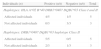
Haplotypes Class I and II in affected and not affected individuals.
| Individuals (n) | Positive (n/t) | Negative (n/t) | Total |
| Haplotypes: HLA A*02 B*40 DRB1*0407 DQB1*03 Class I and II | |||
| Affected individuals | 4/5 | 1/5 | 5 |
| Not affected individuals | 0/3 | 3/3 | 3 |
| Haplotypes: DRB1*0407 DQB1*03 haplotype Class II | |||
| Affected individuals | 5/5 | 0/5 | 5 |
| Not affected individuals | 2/3 | 1/3 | 3 |
The statistical analysis, between what was observed and what was expected: OR=4.0, p=0.0000091.
We did not observe any statistical significance when it was analyzed DRB1*0407: DQB1*03 haplotype OR=1.52, χ2=2.65 and p=0.103.
In four of the members of the family diagnosed with SLE we observed the presence of the HLA A*02 B*40 DRB1*04:07 DQB1*03:02 haplotype – of paternal line (with SLE). Three of the healthy members and the female patient with AIHA did not carry this haplotype.
Our results show the presence of Class II haplotype DRB1*04:07, DQB1*03.02, however did not find a direct association as would be expected because DRB1*04:07 allele is found with high frequency in Amerindian population. However, several authors report this allele in association to different AID. Gonzalez-Treviño et al.6 found that Hashimoto tiroiditis was associated with homozygosity for HLA-DRB1*04, Asenjo et al.,12 found association with DRB1*0407, DQB1*0302 in a Mapuche family with high incidence of Type I Diabetes. Lopez-Tello et al.13 found a significant increase in the frequency of the HLA-DR4 (p=0.16) and the HLA-DR16 (p=0.005) alleles in a Mexican population with SLE.
In this family, we analyzed Class I and Class II haplotype, and all patients with SLE show HLA A*02 B*40 DRB1*0407 DQB1*0302 with high statistical significance. Referring to ours findings, we must consider how HLA Class I in linkage disequilibrium with HLA Class II may be involved in susceptibility or in the development of SLE.
There are some reports showing association in autoimmune disease with HLA Class I alleles, such as Diabetes Mellitus Type I with HLA-A24, B18, -B39-B62-B60.14 The HLA-B60 antigen corresponds to HLA-B*40 which was found in the mestizo family studied associated to SLE. Uchanska-Ziegler B et al.9 found HLA-B*27:05, HLA-A*03:01 or HLA-A*11:01 associated with diseases suspected to have an autoimmune etiology, and postulate that the products of these alleles, due to their unusual ability to bind with high affinity to a particular peptide set during positive selection in the thymus, and involved in shaping an abnormal T cell repertoire which predisposes to acquisition of autoimmune diseases.
HLA-B27 increases the risk of developing ankylosing spondylitis. It is uncertain how HLA-B27 causes this increased risk. Researchers speculate that HLA-B27 may abnormally display peptides that trigger an immune reaction, resulting in the inflammatory process that causes arthritis.8
On the other hand, this forces us to think about the mechanisms by which HLA Class I could contribute to the pathogenesis of this disease, probably the interaction HLA Class I and KIR receptors, play an important role, as has been demonstrated in some autoimmune pathologies.15
SLE shows a strong familial aggregation, with high frequency among first degree relatives of patients. Moreover, in extended families, SLE may coexist with other organ specific autoimmune diseases such as hemolytic anemia, autoimmune thrombocytopenic purpura and thyroiditis.
The primary function of HLA Class I molecules is to present peptide from cleavage of native antigens in the cytosol to CD8+ cells. The conformational differences between alleles of HLA Class I may create variability in the peptide binding properties for presentation to CD8+ cells resulting in the induction of tolerance or self-reactivity. While this could be one of the mechanisms in the HLA Class I intervention in autoimmune diseases, we can not establish whether the same may be one of the factors in the development of lupus and not Class II itself as found in some autoimmune diseases in mestizo population. In reference to the mestizo family studied, an extensive study in this population should be conducted to establish the true participation of HLA Class I region.
Ethical disclosuresProtection of human and animal subjects. The authors declare that the procedures followed were in accordance with the regulations of the responsible Clinical Research Ethics Committee and in accordance with those of the World Medical Association and the Helsinki Declaration.Confidentiality of data. The authors declare that no patient data appears in this article.Right to privacy and informed consent. The authors declare that no patient data appears in this article.
Conflict of interestAuthors declare that they have no conflict of interest.
To director of the J.C. Perrando Hospital in facilitate this study and report that this manuscript was conducted without external funding. Translation: This document was translated by Carla Rolón Martínez.
This research was presented at First French-Argentine Immunology Congress (FAIC 2010), November 2–5, 2010, Buenos Aires, Argentina.


