


We describe a case about a 79-year-old woman with rheumatoid arthritis with chronic left olecranon bursitis refractory to conventional therapy. Following appropriate diagnostic workup, Prototheca spp alga was identified as the causative agent. The exceptional nature of the case is emphasized.
Describimos el caso de una mujer de 79 años con artritis reumatoide que consultó por bursitis crónica de codo izquierdo refractaria a terapia convencional. Tras los estudios de extensión pertinentes se identificó un alga del género Prototheca como agente causal. Se destaca la excepcionalidad del caso.
The genus Prototheca (P) comprises microscopic aerobic algae that do not produce chlorophyll. They are characterised as Gram-positive microorganisms that are difficult to stain and are widely distributed, particularly in humid environments (aquatic environments, crop contamination, trees, etc.).1,2
Infection by this type of microorganism is rare. The first human infection attributed to this species was described by Davies et al. in 1964, and since then, approximately 150 cases have been published in the literature.3
Several species have been described, including P. zopfii, P. filamenta, P. moriformis, and P. stagnora, but P. wikerhamii is the most commonly associated with infections in humans (protothecosis), particularly in the form of opportunistic infections of the skin and soft tissues. The risk is significantly higher in immunocompromised patients, in whom disseminated abdominal forms have been observed in patients undergoing peritoneal dialysis, or haematogenous dissemination in people with central lines.1,3
The pathogenesis of these processes is not fully understood. It has been suggested that transmission occurs through contact with contaminated areas or direct inoculation through traumatic events, surgical interventions, or invasive procedures. Dysfunction of neutrophils and natural killer cells has been suggested as a predisposing factor in patients suffering from these infections.4
At the joint level, it typically behaves like chronic olecranon bursitis. Its predilection for this location has been linked to a predisposition to repeated local trauma.1,5,6 Treatment is controversial given the limited available evidence. Antifungals appear to be effective, particularly amphotericin B and azoles. However, medical treatment is often insufficient and usually requires local surgical intervention.5,7
Clinical observationA 79-year-old woman under follow-up for seropositive, ACPA-negative, erosive rheumatoid arthritis with a 30-year history, currently in clinical remission and under treatment with tocilizumab.
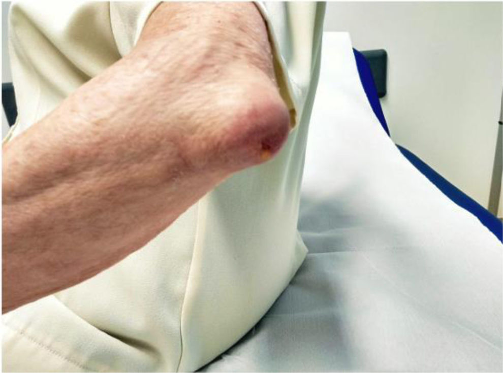
She presented with recurrent pain and swelling of the left olecranon bursa over a period of 6 mo. She was assessed at a private centre where local infiltration was performed on two occasions and oral antibiotic treatment was administered without clinical resolution. The patient denied any history of trauma or a local entry point (Fig. 1).
On examination, signs of bursitis with a small active fistulous orifice were evident. Conventional radiological studies did not reveal images of interest.
Ultrasound-guided drainage was performed, with 2 cm3 of inflammatory-looking fluid being drained. Urgent Gram-negative results were obtained, with no bacterial growth observed in the sample sent.
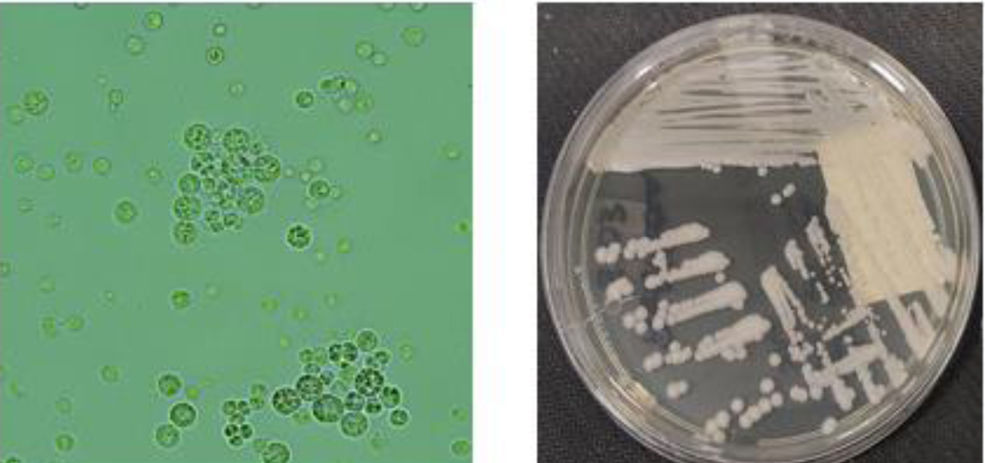
Despite this, due to the high suspicion of an infectious aetiology, local surgery was performed, with persistent signs of bursitis present nine months later. The Microbiology Department re-evaluated the culture (Fig. 2) and identified Prototheca wickerhamii, indicating the need for a new bursectomy with resolution of clinical symptoms.
Images courtesy of the microbiology department of the HUCA. On the left is a direct microscopic examination showing microorganisms in a characteristic "morula" or "spoke-and-wheel" arrangement. On the right is the growth of the bursal fluid sample in Sabouraud culture medium (typically used as a fungal enrichment medium).
Following a thorough medical history review, it was suggested that the patient's regular river swimming could be the source of contamination for the symptoms presented.
DiscussionProtothechosis is a rare clinical condition, although it may be a reason for referral to rheumatology clinics due to the profile of the affected patients (mainly immunocompromised) and the possible musculoskeletal presentation (chronic olecranon bursitis.
Although certain antifungal agents, such as amphotericin B, have proven effective in treating the condition, a surgical approach is often necessary for adequate resolution.
ConclusionProtothecosis should be considered in the differential diagnosis of chronic olecranon bursitis that does not respond to conventional treatment and/or antibiotics. This suspicion should be communicated to microbiology services.
The authors have no conflict of interests to declare.