Journal Information
Vol. 11. Issue 3.
Pages 179-181 (May - June 2015)
Vol. 11. Issue 3.
Pages 179-181 (May - June 2015)
Images in Clinical Rheumatology
Full text access
Giant Baker’ Cyst. Differential Diagnosis of Deep Vein Thrombosis
Quiste de Baker gigante. Diagnóstico diferencial de trombosis venosa profunda
Visits
14752
Noelia Alonso-Gómez
Corresponding author
nelinew@hotmail.com

Corresponding author.
, Antonio Pérez-Piqueras, Antonio Martínez-Izquierdo, Felipe Sáinz-González
Angiología y Cirugía Vascular, Hospital Central de la Defensa «Gómez-Ulla», Madrid, Spain
This item has received
Article information
Full Text
Bibliography
Download PDF
Statistics
Figures (5)
Show moreShow less
Full Text

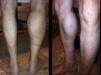
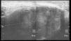
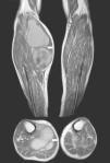
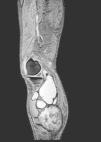
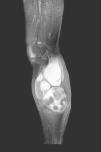
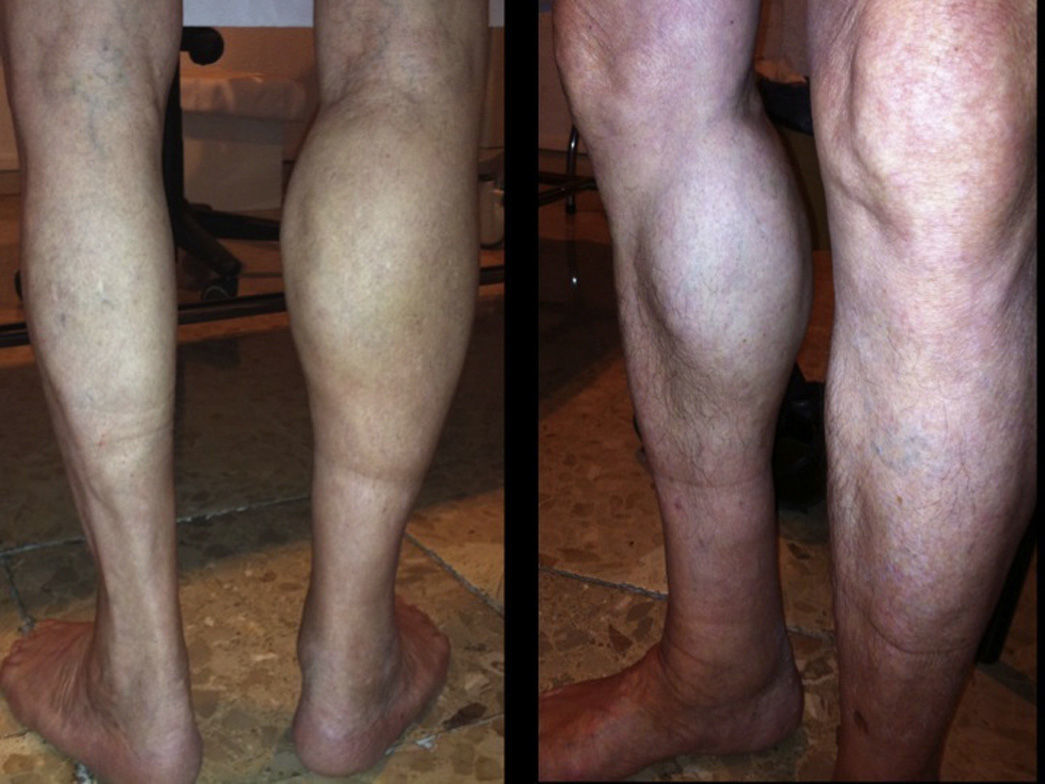
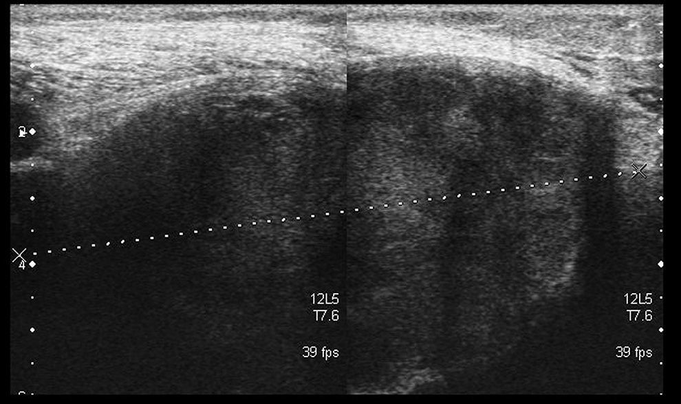
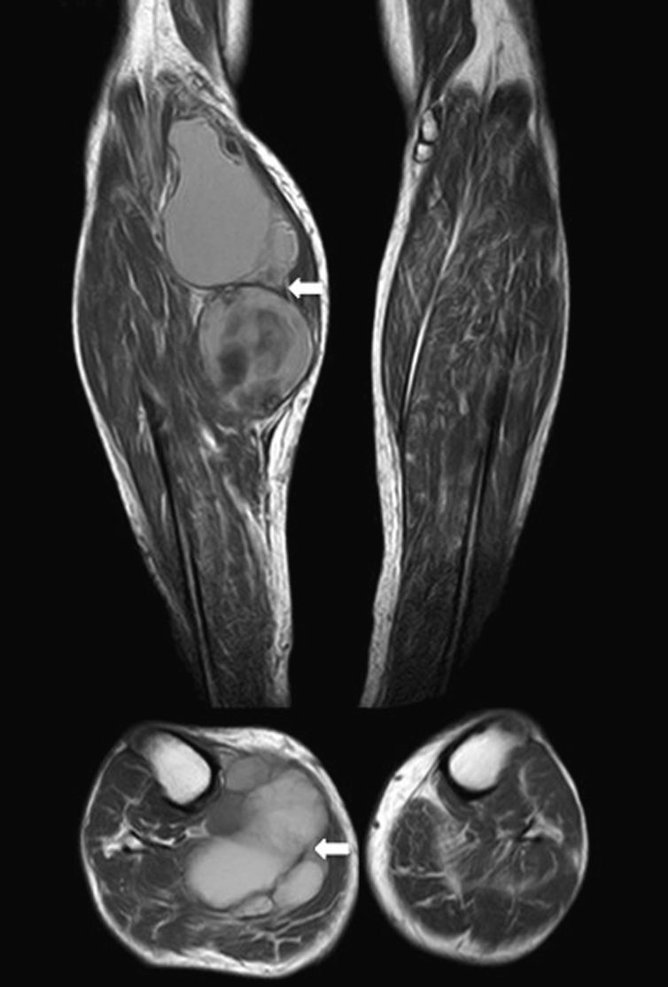
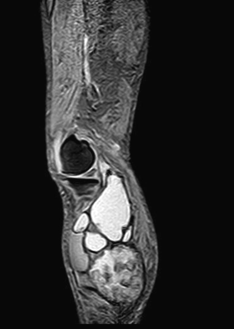
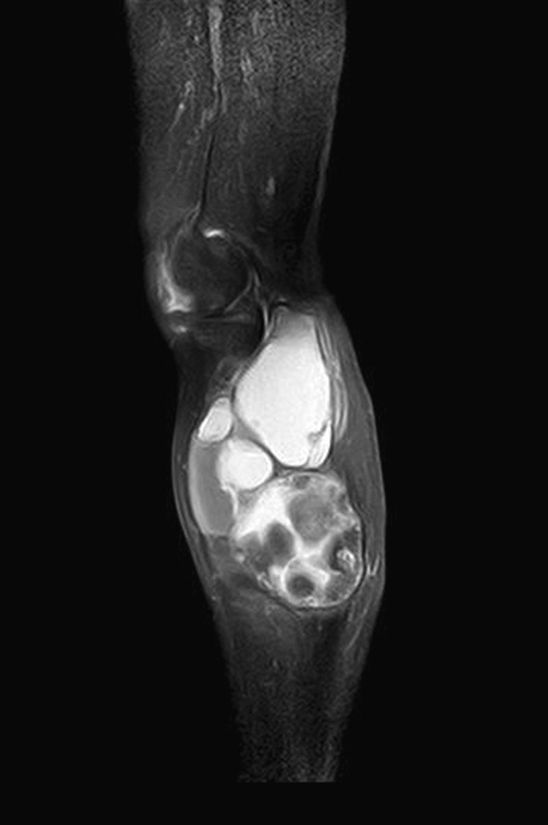
We present the case of an 83-year-old male patient with osteoarthritis, who came to the emergency room due to acute inflammation of the leg simulating DVT. On physical examination, there was a mass of elastic consistency, non-pulsatile, painful, in the popliteal fossa, extending to the leg (Fig. 1). The differential diagnosis was made with Baker cyst. The patient underwent, as a technique of choice, an ultrasound. The color Doppler mode showed patency of the deep venous system. Musculoskeletal ultrasound evidenced a well-defined collection, adjacent to the medial gastrocnemius muscle belly, full of vague echoes predominantly of low amplitude (Fig. 2) Despite being frequent, the size of the tumor made ultrasound insufficient and we decided to perform specific knee MRI with T1 potentiated sequences, T2* sequences (Figs. 3–5) and, after administration of intravenous contrast, we confirmed the existence of giant popliteal cyst of 17cm×9cm. The patient was treated conservatively, using, in the first place, nonsteroidal anti-inflammatory drugs, with clinical improvement and asymptomatic after the first 6 months. For this reason, along with the age of the patient and in spite of the cyst barely changing its size, surgical treatment has so far been dismissed.

Fig. 1.

Gastrocnemius asymmetry.

(0.12MB).
Fig. 2.

Ultrasound. Transversal view. Heterogeneous popliteal cyst. Appearance corresponding to fibrinous blood clot.

(0.1MB).
Fig. 3.

MR. Axial and coronal T1-weighted images. Well circumscribed mass (arrows). Increased intensity signal compatible with a subacute presence of blood products.

(0.08MB).
Fig. 4.

MR. Sagittal STIR image. The hematoma shows diffuse increased signal intensity.

(0.07MB).
Fig. 5.

MR. Sagittal T1-weighted image with fat saturation. Peripheral potentiation is observed compared to the precontrast image but no significant image central enhancement, indicating its cystic nature.

(0.05MB).

Complicated Baker's cyst is a very common entity. The prevalence increases with age. However, we have only found one published case1 of similar dimensions. Cysts containing over150ml produce neurovascular compression phenomena at the popliteal level, being associated with2,3 other conditions, such as rheumatoid arthritis.4,5 Both for possessing a broad spectrum of presentations and the characteristics of the synovial liquid seen by ultrasound sometimes makes this a diagnostic challenge.6,7

Ethical ResponsibilitiesProtection of people and animals

The authors declare this research did not perform experiments on humans or animals.

Data confidentiality

The authors declare that they have followed the protocols of their workplace regarding the publication of patient data.

Right to privacy and informed consent

The authors have obtained the informed consent of patients and/or subjects referred to in the article. This document is in the possession of the corresponding author.

Conflict of Interest

The authors have no conflict of interest to state.

References
[1]
S. Garg, T. Al-Jabri, S. Mutnal, F. Moftah.
A giant ganglion cyst of the semimembranous tendon: a case report.
[2]
J.E. Sanchez, N. Conkling, N. Labropoulos.
Compression syndromes of the popliteal neurovascular bundle due to Baker cyst.
J Vasc Surg, 54 (2011), pp. 1821-1829
[3]
R. Rettori, O. Boespflug.
Popliteal vein entrapment, popliteal cyst, desmoid tumor and fabella syndrome.
J Mal Vasc, 15 (1990), pp. 182-187
[4]
F. Hoffmann-González, C. Hernández-Díaz, C. Solano-Ávila, A.G. López-Reyes, A. Peña-Ayala, C. Pineda-Villaseñor.
Giant Baker's cyst treated with intralessional methotrexate.
Cir Cir, 81 (2013), pp. 64-68
[5]
J. Ravlić-Gulan, G. Gulan, S. Novak, B. Sestan.
Rapid recurrence of a giant popliteal cyst in a patient with rheumatoid arthritis.
J Clin Rheumatol, 15 (2009), pp. 300-302
[6]
W.C. Torreggiani, K. Al-Ismail, P.L. Munk, C. Roche, C. Keogh, S. Nicolaou, et al.
The imaging spectrum of Baker's (popliteal) cysts.
Clin Radiol, 57 (2002), pp. 681-691
[7]
E.E. Ward, J.A. Jacobson, D.P. Fessell, C.W. Hayes, M. van Holsbeeck.
Sonographic detection of Baker's cysts: comparison with MR imaging.
Am J Roentgenol, 176 (2001), pp. 373-380

Please cite this article as: Alonso-Gómez N, Pérez-Piqueras A, Martínez-Izquierdo A, Sáinz-González F. Quiste de Baker gigante. Diagnóstico diferencial de trombosis venosa profunda. Reumatol Clin. 2015;11:179–181.

Copyright © 2014. Elsevier España, S.L.U.. All rights reserved
Idiomas
Reumatología Clínica (English Edition)
Article options
Tools
es en

¿Es usted profesional sanitario apto para prescribir o dispensar medicamentos?

Are you a health professional able to prescribe or dispense drugs?